
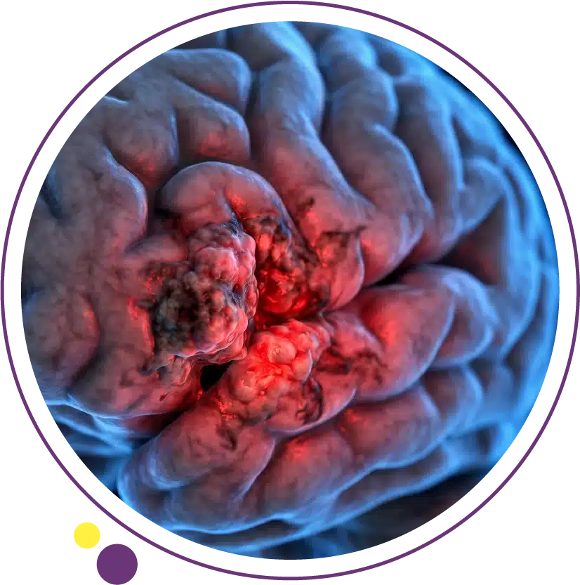
Glioblastoma is the most common and most aggressive primary brain tumor in adults. It grows quickly, infiltrates surrounding brain tissue, and produces symptoms that can escalate from mild to severe within weeks. Despite decades of intensive research and numerous clinical trials, it remains one of the most challenging cancers to treat. Understanding what glioblastoma is, how it is diagnosed, what treatment involves, and what outcomes to realistically expect is essential for patients and families facing this diagnosis.
This article covers glioblastoma comprehensively: its biology and why it is so aggressive, the symptoms it produces and why they vary by tumor location, how MRI and molecular testing establish the diagnosis, the Stupp protocol and other treatment approaches, emerging therapies, and the central role that serial brain MRI plays throughout the entire treatment and monitoring journey.
What Is Glioblastoma?
Glioblastoma, formally classified as glioblastoma multiforme (GBM) or WHO Grade 4 astrocytoma, is a malignant glial tumor arising from astrocytes, the star-shaped support cells of the brain. Under the updated 2021 WHO Classification of Central Nervous System Tumours, glioblastoma is defined specifically as an IDH-wildtype diffuse astrocytic tumor with certain molecular characteristics, distinguishing it from IDH-mutant high-grade gliomas which have a significantly better prognosis. Glioblastoma represents approximately fifteen percent of all primary brain tumors and approximately forty-five percent to fifty percent of all malignant brain tumors.
Glioblastoma can arise de novo (primary GBM), which is the most common form, typically affecting patients over fifty and progressing rapidly from the outset. Less commonly it arises through malignant progression from a lower-grade glioma (secondary GBM), which tends to affect younger patients and has somewhat better biological behaviour. Both forms produce the same imaging appearance and are managed similarly, though molecular profiling distinguishes them. At Images Diagnostic Center in Kuwait, 3 Tesla MRI with contrast provides the imaging quality needed for the initial characterisation and the serial monitoring that GBM management requires throughout treatment.
Why Glioblastoma Is So Aggressive
Several biological features make glioblastoma particularly resistant to treatment. First, it is diffusely infiltrative: tumor cells spread along white matter tracts far beyond the visible enhancing mass, making complete surgical removal impossible. The cells that remain after surgery eventually drive recurrence. Second, glioblastoma is genetically heterogeneous, meaning different regions of the same tumor contain cells with different genetic profiles. This intratumoral heterogeneity means that a treatment targeting one genetic variant may be ineffective against others. Third, glioblastoma exploits the blood-brain barrier selectively: the tumor core breaks down the barrier, which is why contrast enhancement is visible on MRI, but the infiltrating margin retains enough barrier function to exclude many systemic chemotherapy drugs.
Fourth, glioblastoma creates an immunosuppressive microenvironment that shields it from immune attack, which partially explains why immunotherapy approaches that have transformed treatment in lung and skin cancer have been less successful in GBM to date. Understanding these biological obstacles is essential context for appreciating why even the best current treatments achieve limited survival extension. The MRI service at Images tracks the local and systemic consequences of these biological processes throughout treatment.
Glioblastoma Symptoms
Glioblastoma symptoms typically develop over days to weeks rather than the months typical of lower-grade gliomas, reflecting the rapid growth rate of the tumor. The most common presenting symptoms include progressive headache that is often worst in the morning and worsened by lying flat, new-onset seizures in an adult with no prior seizure history, focal neurological deficits such as limb weakness, visual disturbance, or speech difficulty that correspond to the functional brain region where the tumor is growing, and cognitive changes including memory impairment, confusion, and difficulty with complex tasks.
Personality and behaviour changes are a particularly important early sign that is often attributed initially to stress, depression, or another psychiatric cause rather than a neurological one. Frontal lobe glioblastomas are especially likely to produce these changes because of the frontal lobe role in executive function and social behaviour. A previously reliable, organised individual who suddenly becomes disinhibited, apathetic, or cognitively impaired over a period of weeks has a symptom pattern that demands brain imaging. For a full breakdown of how brain tumor symptoms relate to anatomical location, our article on brain tumor symptoms covers this in comprehensive clinical detail. Brain MRI at Images is the investigation of choice when these presentations occur.
How Glioblastoma Is Diagnosed
MRI: The Essential Diagnostic Study
MRI with gadolinium contrast is the imaging modality of choice for glioblastoma diagnosis and characterisation. On MRI, glioblastoma typically appears as an irregular, heterogeneously enhancing mass with a central area of necrosis that does not enhance, surrounded by an enhancing rim of viable tumor, which is itself surrounded by extensive non-enhancing infiltration visible on FLAIR sequences. This ring-enhancing pattern with central necrosis in a supratentorial location in an older adult is highly characteristic of glioblastoma, though it can occasionally be mimicked by brain metastasis or abscess.
Advanced MRI techniques add further diagnostic precision. MR spectroscopy demonstrates elevated choline and reduced NAA consistent with high-grade malignancy. Perfusion MRI shows elevated cerebral blood volume in the tumor consistent with high-grade neovascularisation. Diffusion-weighted imaging assesses cellularity and can help distinguish different tumor regions. The 3 Tesla MRI at Images provides the field strength that maximises the quality of all of these sequences, offering neurosurgeons and neuro-oncologists the detailed pre-operative imaging they need for treatment planning. For patients who experience anxiety in enclosed scanners, Open MRI is available as an alternative.
Biopsy and Molecular Profiling
MRI findings, however characteristic, do not replace histopathological diagnosis. A tissue sample obtained through stereotactic biopsy or at the time of surgical resection is essential to confirm the diagnosis, determine the WHO grade, and provide material for molecular profiling. The molecular profile of a glioblastoma is now as clinically important as its histological grade. IDH mutation status is the most critical biomarker: IDH-wildtype tumors are glioblastoma by WHO 2021 definition and carry the worse prognosis, while IDH-mutant high-grade gliomas have a meaningfully better prognosis despite sharing some histological features.
MGMT promoter methylation status is the next most clinically important molecular marker, predicting benefit from temozolomide chemotherapy. MGMT-methylated glioblastomas, which account for approximately forty to fifty percent of cases, respond better to temozolomide and have modestly better survival compared to unmethylated tumors. TERT promoter mutations, EGFR amplification, and chromosome 10q deletion are additional molecular markers with prognostic and potentially predictive significance. The pre-biopsy MRI from Images provides the roadmap that neurosurgeons use to identify the optimal biopsy target within the tumor mass. The CT service at Images may complement this with stereotactic planning support.
Staging and Extent of Disease Assessment
Unlike most systemic cancers, glioblastoma staging is not formally described using a TNM system because distant metastasis outside the central nervous system is extremely rare. Clinical staging of glioblastoma focuses instead on local disease extent and functional impact. The key imaging question is not whether the tumor has spread to distant organs but rather how much of the eloquent brain it involves and whether the involvement patterns preclude safe surgical debulking. Surgeons use preoperative MRI to assess the relationship of the tumor to motor cortex, language areas, visual pathways, and major vascular structures.
Multi-sequence preoperative MRI including functional MRI (fMRI) to map eloquent cortex and diffusion tensor imaging (DTI) to visualise white matter tracts may be performed at specialist neurosurgical centres before resection of tumors in eloquent brain locations. This advanced planning maximises the extent of safe surgical resection while minimising the risk of new permanent neurological deficits. The standard preoperative brain MRI at Images provides the anatomical baseline that begins this assessment. For patients interested in understanding causes and risk factors for their diagnosis, our article on brain tumor causes addresses the most common questions.
Treatment: The Stupp Protocol
The standard first-line treatment for glioblastoma was established by the landmark Stupp trial published in 2005 and remains the backbone of treatment today. The Stupp protocol consists of maximal safe surgical resection followed by concurrent temozolomide (TMZ) chemotherapy administered daily during six weeks of fractionated radiation to the tumor bed, followed by six cycles of adjuvant TMZ administered monthly for five days per cycle. The Stupp protocol demonstrated a median survival of fourteen to fifteen months compared to approximately twelve months with radiation alone, and a meaningful improvement in two-year survival from eleven percent to twenty-seven percent.
Surgical resection serves multiple purposes: it reduces the tumor burden that subsequent treatment must address, provides tissue for histological and molecular diagnosis, relieves mass effect and symptom burden, and may provide independent survival benefit when maximal safe resection is achieved. Gross total resection, when safely achievable, is associated with longer progression-free and overall survival compared to subtotal resection or biopsy alone. Brain MRI within twenty-four to seventy-two hours after surgery is performed to assess the extent of resection before post-operative oedema distorts the imaging. The post-operative MRI service at Images supports this critical assessment in Kuwait. The full imaging services at Images then continue throughout the chemoradiation and adjuvant chemotherapy phases to monitor treatment response.
Bevacizumab, Tumor Treating Fields, and Emerging Therapies
Bevacizumab, an anti-VEGF antibody that inhibits tumor angiogenesis, is approved for recurrent glioblastoma based on its ability to produce radiographic response and extend progression-free survival. Its impact on overall survival in recurrent GBM has been more modest, and it remains a standard option for patients with symptomatic recurrence rather than a curative treatment. Bevacizumab use also produces a specific imaging effect called pseudoresponse, where the enhancement appears to dramatically decrease due to restored blood-brain barrier integrity rather than true tumor reduction, which complicates MRI response assessment at this treatment stage.
Tumor treating fields (TTFields), delivered through a wearable device that applies alternating electric fields to the scalp, have shown survival benefit in newly diagnosed MGMT-unmethylated glioblastoma when added to maintenance temozolomide, leading to regulatory approval in several countries. Immunotherapy approaches including checkpoint inhibitors, CAR-T cells, and vaccine-based strategies are under active investigation in multiple clinical trials but have not yet produced the breakthroughs seen in other cancer types. Targeted therapy with specific inhibitors for EGFR amplification, FGFR alterations, and other molecular drivers is also being explored. Serial MRI monitoring throughout these treatments is essential for assessing response and distinguishing true progression from pseudoprogression, which is a treatment-related imaging change that can appear as apparent tumor growth in the weeks to months after chemoradiation. The MRI team at Images provides this critical ongoing monitoring in Kuwait.
Prognosis and Survival
Glioblastoma remains one of the most challenging cancers to treat, with a median overall survival of approximately fifteen to eighteen months from diagnosis for patients treated with the Stupp protocol. Two-year survival is achieved by approximately twenty-five to thirty percent of patients, and five-year survival by fewer than ten percent. These statistics reflect the population average; individual patients vary considerably based on age at diagnosis, performance status, extent of surgical resection, MGMT methylation status, and other molecular features. Younger patients with good functional status, high MGMT methylation, and maximal surgical resection represent the subgroup with the most favourable outcomes.
It is important for patients and families to understand that these statistics are based on historical cohorts and that the outcomes landscape is slowly improving with advances in molecular-guided treatment selection and supportive care. Clinical trial participation is an option for eligible patients at diagnosis and at recurrence and may provide access to emerging therapies not yet available outside the trial setting. Palliative and supportive care services are an essential component of glioblastoma management from diagnosis onward, addressing symptoms, maintaining quality of life, and supporting families alongside the active anti-tumor treatment pathway. Serial CT and MRI imaging at Images supports every stage of this care pathway in Kuwait. For complementary context on glioma as a category and how GBM fits within it, our article on glioma symptoms explains how different glioma grades present differently and how imaging helps distinguish them.
Frequently Asked Questions
Is glioblastoma always fatal?
Glioblastoma is not curable with current treatments in the vast majority of patients, and median survival is approximately fifteen to eighteen months with standard treatment. However, a meaningful minority of patients survive well beyond two years, and a smaller proportion achieve long-term survival exceeding five years. Favourable prognostic factors including younger age, good performance status, MGMT methylation, and maximal safe resection are associated with the best outcomes. Clinical trial participation may offer access to emerging therapies that improve on current standard results.
What is the difference between GBM and other gliomas?
Glioblastoma is a WHO Grade 4 astrocytoma, specifically defined as IDH-wildtype with certain molecular characteristics. Lower-grade gliomas (Grades 1 to 3) grow more slowly, produce symptoms more gradually, and carry a better prognosis. IDH-mutant Grade 4 astrocytomas have a significantly better prognosis than IDH-wildtype glioblastoma despite sharing some histological features. The molecular distinction is now as important as the microscopic appearance for both diagnosis and treatment planning.
How quickly does glioblastoma grow?
Glioblastoma grows rapidly, with imaging studies showing volume doubling times of weeks to a few months. This rapid growth explains the short interval between first symptoms and significant neurological disability when diagnosis is delayed. The infiltrative growth pattern means that tumor cells extend well beyond the visible MRI abnormality, which is why even gross total resection of the visible tumor does not eliminate all tumor cells. Follow-up MRI is performed every two to three months during treatment to detect early progression.
What is pseudoprogression in glioblastoma?
Pseudoprogression is an imaging phenomenon where the MRI appears to show increased tumor enhancement and surrounding oedema in the weeks to months after completing chemoradiation, mimicking true tumor growth when in reality the change reflects treatment-related inflammation and tissue reaction rather than actual cancer progression. It occurs in approximately thirty to forty percent of patients following the Stupp protocol and is more common in MGMT-methylated tumors. Distinguishing pseudoprogression from true progression requires advanced MRI techniques, clinical assessment, and often serial follow-up imaging to observe subsequent behaviour over time.
Where can I arrange glioblastoma-related brain MRI in Kuwait?
Images Diagnostic Center provides 3 Tesla brain MRI across three Kuwait branches, supporting all stages of glioblastoma imaging from initial diagnosis through pre-operative planning, post-operative assessment, treatment response monitoring, and surveillance for recurrence. You can contact Images to arrange the appropriate study once your neurosurgical or neuro-oncology team has made a referral.
Navigating Glioblastoma With Accurate Information and Expert Imaging
A glioblastoma diagnosis is one of the most difficult news a patient and family can receive, and the clinical journey that follows requires both expert medical care and high-quality imaging support at every stage. From the initial MRI that raises the diagnostic concern, through the pre-surgical imaging that guides the operation, to the serial scans that track treatment response and detect recurrence, imaging is the constant companion of every major decision in GBM management. Images Diagnostic Center provides 3 Tesla brain MRI across three Kuwait branches to support every step of this pathway.
To arrange brain MRI for glioblastoma evaluation or monitoring, contact Images directly.