
Glioma is not a single disease. It is an umbrella term for a diverse family of brain tumors arising from glial cells, the supportive cells of the central nervous system. The symptoms a glioma produces vary dramatically depending on two factors: the grade of the tumor, which determines how rapidly it grows and how aggressively it invades surrounding brain tissue, and the location within the brain, which determines which specific neurological functions are disrupted. A low-grade glioma in the frontal lobe of a thirty-year-old may produce only seizures for years, while a high-grade glioma in the same location may cause rapid cognitive decline within weeks.
This article explains glioma symptoms by tumor type and grade, addresses the personality shifts and memory difficulties that are among the most commonly missed early signs, describes how location within the brain shapes the specific symptom profile, and explains why brain MRI is the definitive next step when any of these symptom patterns develops.
What Is a Glioma?
Gliomas arise from different types of glial cells: astrocytes give rise to astrocytomas and glioblastoma, oligodendrocytes give rise to oligodendrogliomas, and ependymal cells give rise to ependymomas. Under the 2021 WHO Classification of CNS Tumours, gliomas are graded from 1 to 4 based on histological and molecular features, with Grade 1 being the most benign and Grade 4 (glioblastoma) being the most malignant. IDH mutation status has become a defining molecular marker that divides diffuse gliomas into two major biological groups with fundamentally different prognoses: IDH-mutant gliomas (which are generally lower grade and carry better prognoses) and IDH-wildtype glioblastoma (the most aggressive form).
For patients and families, understanding glioma grade matters because it directly predicts how quickly symptoms will develop and progress, how the tumor will appear on MRI, and what treatment options are available. At Images Diagnostic Center in Kuwait, 3 Tesla MRI with contrast provides the imaging quality needed to characterise gliomas and differentiate between grades based on enhancement patterns, perfusion characteristics, and other advanced imaging features. For a detailed discussion of glioblastoma specifically, our comprehensive article on glioblastoma covers the full clinical picture from diagnosis through treatment.
Low-Grade Glioma Symptoms: Subtle and Easy to Miss
Seizures as the Most Common First Presentation
New-onset seizures in an adult are the most common first presentation of low-grade glioma, accounting for sixty to ninety percent of initial presentations in Grade 2 tumors. Because low-grade gliomas grow slowly and are less likely to produce significant mass effect or disrupt the blood-brain barrier in their early stages, they may not cause headaches or visible neurological deficits for years. Instead, the irritative effect of the tumor on surrounding cortical tissue triggers abnormal electrical activity that produces focal or generalised seizures. The seizure may be the first indication that anything is wrong.
Focal seizures from low-grade gliomas may take many forms depending on the tumor location: rhythmic jerking of one limb, transient sensory disturbances such as tingling or numbness in one area, visual symptoms including flashing lights or formed visual hallucinations, olfactory hallucinations (smelling an odour that is not present), or brief episodes of altered awareness with automatic behaviours. Any new-onset seizure in an adult who has never had a seizure before requires brain imaging. MRI at Images is the investigation of choice, with contrast enhancement used to assess blood-brain barrier integrity and help distinguish low-grade from high-grade disease.
Subtle Cognitive and Personality Changes in Low-Grade Glioma
Beyond seizures, low-grade gliomas produce subtle neurological changes that are easily attributed to other causes. Mild difficulty with word-finding or language processing, slightly impaired memory, reduced concentration, or vague changes in personality and social behaviour are all consistent with a slowly growing glioma in a functionally relevant brain region. These changes are gradual enough that the patient and people around them may not notice them clearly until they are compared against how the person functioned several years earlier.
In low-grade gliomas specifically, the long natural history means that subtle symptoms may be present for months or even years before the seizure or other more obvious event that leads to MRI and diagnosis. Retrospective histories from patients and families often reveal that mild changes in personality, word-finding, or mood had been developing quietly for an extended period before the diagnosis was made. The brain MRI service at Images is the investigation that converts clinical suspicion into diagnostic certainty in these presentations. Our article on brain tumor symptoms covers the full clinical range of presentations that indicate a need for urgent brain evaluation.
High-Grade Glioma Symptoms: Rapid and Progressive
Rapid-Onset Headaches
High-grade gliomas, including Grade 3 anaplastic gliomas and Grade 4 glioblastoma, grow rapidly enough to produce significant mass effect within weeks. This raised intracranial pressure manifests as headaches that are characteristically worse in the morning, worse when lying flat, worsened by the Valsalva manoeuvre (coughing, straining), and often accompanied by nausea or vomiting. These features reflect the postural variation in intracranial pressure that occurs with changes from upright to supine position. A new headache with any of these characteristics in a patient over forty should prompt brain imaging as a priority rather than empirical headache treatment without investigation.
The headache from high-grade glioma is progressive over days to weeks rather than the episodic or fluctuating pattern of migraine or tension headache. It does not respond to standard analgesia in the sustained way that tension headache does, and it is typically accompanied by at least one other neurological symptom when evaluated carefully. CT of the brain may be arranged urgently to exclude haemorrhage or severe mass effect, followed by MRI for full characterisation. Both are available at Images in Kuwait.
Focal Neurological Deficits
High-grade gliomas produce focal neurological deficits that reflect the specific brain region invaded by the tumor. Motor cortex involvement produces contralateral limb weakness that may begin as subtle clumsiness with fine motor tasks and progress to significant arm or leg weakness. Language area involvement in the dominant hemisphere produces expressive or receptive aphasia. Parietal lobe involvement causes contralateral sensory loss, spatial disorientation, or neglect of one side of space. Occipital involvement causes contralateral visual field loss. These deficits develop progressively over days to weeks rather than the instantaneous onset characteristic of stroke, which is an important clinical distinguishing feature.
Sudden focal neurological deficit in a patient with a known or suspected glioma may indicate haemorrhage into the tumor, which is a neurological emergency requiring immediate CT brain. For patients without a prior diagnosis, progressive focal neurological deficit over days to weeks represents an urgent indication for brain MRI. The 3 Tesla MRI service at Images provides the anatomical detail needed to characterise these focal lesions precisely and determine their relationship to critical brain structures before treatment planning. For patients who have difficulty with enclosed spaces, Open MRI is also available.
Personality Shifts and Memory Trouble: The Signs Most Often Missed
Personality change and memory impairment deserve special attention because they are the glioma symptoms most frequently attributed to other causes, most commonly depression, anxiety, burnout, or early dementia, before the neurological origin is recognised. A patient who becomes uncharacteristically irritable, disinhibited, apathetic, or socially inappropriate; who makes impulsive decisions inconsistent with their normal character; or who begins to lose track of appointments, conversations, and daily tasks may be experiencing the early effects of a frontal or temporal lobe glioma on executive and memory function.
These changes are often first noticed by family members, colleagues, or close friends rather than by the patient, who may lack insight into their own changed behaviour when the frontal lobe is affected. A psychiatric referral for new-onset behavioural disturbance in a middle-aged or older adult without prior psychiatric history should routinely be preceded or accompanied by brain imaging to exclude a structural cause. New personality or cognitive changes in the context of any other neurological symptom make the case for brain MRI even more compelling. The full imaging services at Images support this evaluation across all three Kuwait branches. Our article on brain tumor causes provides complementary context on the risk factors that increase the pre-test probability of a brain tumor in this clinical situation.
How IDH Status Relates to Symptom Speed and Progression
IDH mutation status, while a molecular laboratory finding rather than a clinical symptom, has direct implications for the tempo of symptom progression that patients and clinicians observe. IDH-mutant gliomas are typically lower grade and grow more slowly, meaning their symptom progression is correspondingly more gradual. A patient with an IDH-mutant Grade 2 oligodendroglioma may have seizures as the only symptom for years before other neurological changes develop. IDH-wildtype glioblastoma, by contrast, grows rapidly and produces progressive symptoms over weeks rather than years.
This difference in biological tempo has important clinical implications. It means that a patient with an IDH-mutant low-grade glioma presenting with seizures may have reasonable quality of life for an extended period before significant disability develops, whereas a patient with IDH-wildtype glioblastoma at the same symptom stage will typically experience rapid progression. The molecular profile obtained from biopsy material shapes these prognosis conversations alongside the imaging findings. The brain MRI at Images provides the imaging baseline that is correlated with the molecular findings to give the complete clinical picture. For patients and families wanting to understand the glioma spectrum more broadly, the Images health blog provides educational resources on brain tumor topics.
Glioma Symptoms by Brain Location
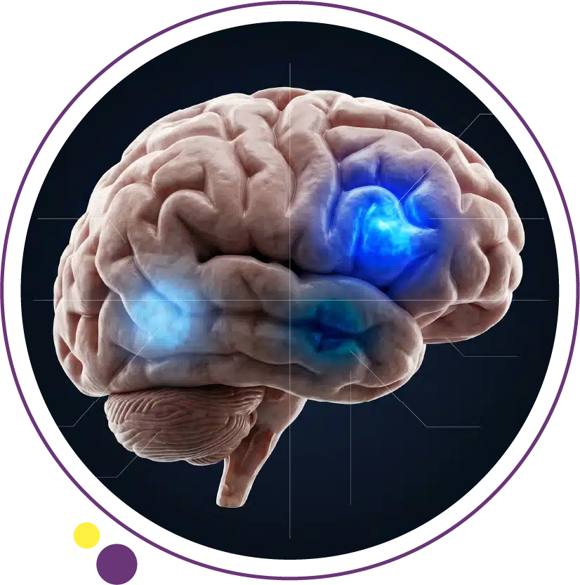
The anatomical location of a glioma shapes the specific symptom profile as precisely as a map shapes the territory it represents. Frontal lobe gliomas produce personality change, disinhibition, apathy, executive dysfunction, difficulty with planning and organisation, and contralateral limb weakness when the motor strip is involved. Temporal lobe gliomas produce seizures (which are particularly common because of the temporal lobe intrinsic excitability), memory impairment, language comprehension difficulty when the dominant hemisphere is involved, and hearing or smell changes.
Parietal lobe gliomas produce contralateral sensory loss, spatial disorientation, difficulty with reading or calculation in the dominant hemisphere, and neglect of the contralateral space in the non-dominant hemisphere. Occipital gliomas primarily produce visual symptoms including contralateral visual field loss and visual hallucinations. Thalamic and deep midline gliomas produce contralateral sensory and motor disturbances combined with raised intracranial pressure symptoms because of their proximity to cerebrospinal fluid pathways. Brainstem gliomas, which are more common in children than adults, produce multiple cranial nerve palsies, ataxia, and long tract signs. This anatomical symptom localisation framework is the same one that guides the interpretation of brain MRI findings at Images, where the imaging report correlates the visible lesion location with the expected clinical symptom territory.
When Glioma Symptoms Require Urgent Evaluation
Any first seizure in an adult requires brain imaging as a priority, ideally brain MRI within twenty-four to forty-eight hours. Progressive focal neurological deficit developing over days to weeks without a clear explanation requires urgent neurological referral and brain MRI. New-onset personality change or cognitive decline of recent onset in a previously healthy middle-aged adult warrants neurological assessment and brain imaging as part of the workup, not as a last resort after other investigations. Rapid progression of any existing neurological symptom, particularly in a patient with a known prior glioma, requires immediate assessment for tumor progression or treatment complication.
Sudden severe neurological deterioration in any patient with a known or suspected brain tumor is an emergency requiring immediate CT brain to exclude haemorrhage into the tumor or acute hydrocephalus. CT at Images provides the fast initial assessment for these emergency presentations, followed by MRI for complete characterisation when the situation is stable. The Images team can arrange priority brain imaging when clinical urgency is indicated.
Frequently Asked Questions
What is the difference between glioma and glioblastoma?
Glioma is the broad category that includes all tumors arising from glial cells, spanning WHO Grades 1 to 4. Glioblastoma (WHO Grade 4, IDH-wildtype) is the most aggressive subtype within this category. All glioblastomas are gliomas, but most gliomas are not glioblastomas. Lower-grade gliomas including pilocytic astrocytoma (Grade 1), diffuse astrocytoma (Grade 2), and oligodendroglioma (Grade 2) have substantially better prognoses than glioblastoma and grow much more slowly.
Why do low-grade gliomas cause seizures so commonly?
Low-grade gliomas preferentially affect the cerebral cortex and have a particular propensity for certain cortical regions including the temporal and frontal lobes. The tumor infiltrates and disrupts cortical circuits without necessarily killing the neurons it involves in early stages, creating areas of abnormal electrical activity that lower the seizure threshold. IDH-mutant gliomas also produce glutamate, which has excitatory neuroactive effects on surrounding tissue. The result is a high prevalence of seizures as the presenting symptom, often before any other neurological deficit is apparent.
Can glioma symptoms be present for years before diagnosis?
Yes, particularly for low-grade IDH-mutant gliomas. Some patients have subtle symptoms including mild cognitive changes or infrequent seizures for two to five years or more before the diagnosis is made. This is partly because the symptoms are mild and attributed to other causes, and partly because the tumors grow slowly enough that the overall neurological function remains relatively preserved for extended periods. In contrast, high-grade gliomas produce symptoms over weeks rather than years due to their rapid growth.
Is CT or MRI better for glioma evaluation?
MRI is substantially superior to CT for glioma evaluation. CT may detect a mass or abnormality that prompts further investigation, but MRI with contrast provides the detail needed to characterise the tumor, assess its grade, define its extent, evaluate blood-brain barrier disruption through enhancement patterns, and plan surgical or radiotherapy targets. Advanced MRI sequences including FLAIR, perfusion, diffusion, and spectroscopy add further diagnostic precision. 3 Tesla MRI at Images provides the field strength needed to maximise the quality of all of these sequences for glioma evaluation in Kuwait.
Are oligodendroglioma symptoms different from astrocytoma symptoms?
Both are low-grade gliomas and share many symptom features. Oligodendrogliomas have a particularly strong predilection for the frontal and temporal lobes and have an even higher rate of seizures as the presenting symptom, sometimes exceeding eighty to ninety percent. Oligodendrogliomas also tend to contain calcifications that may be visible on CT. Astrocytomas have a more variable distribution and slightly lower seizure rate as a presenting feature. The molecular distinction (1p/19q co-deletion in oligodendrogliomas) has greater prognostic significance than the clinical symptom differences between the two subtypes.
The Right Imaging at the Right Time Makes the Difference
Glioma symptoms are as varied as the tumors themselves, ranging from a single focal seizure in an otherwise well young adult to rapid cognitive collapse in an older patient with a high-grade tumor. What unites all these presentations is that brain MRI with contrast is the investigation that provides the definitive answer when clinical suspicion is raised. Acting on the full range of glioma symptoms, including the subtle ones like personality change, memory difficulty, and infrequent seizures, rather than waiting for more obvious deficits to develop, maximises the probability of finding the tumor at a stage when meaningful treatment options are available.
Images Diagnostic Center provides 3 Tesla brain MRI across three Kuwait branches to support glioma evaluation at every stage from initial symptom assessment to treatment monitoring:
To arrange brain MRI for glioma symptom evaluation or monitoring in Kuwait, contact Images directly.