
When a lung cancer diagnosis is confirmed, one of the first questions that follows is: what stage is it? The stage tells the patient and their clinical team how far the cancer has spread, which treatment approaches are available and appropriate, and what the realistic expectations for outcomes are. Stage is not simply a number; it represents the biological state of the disease at the time of diagnosis and is the single most important factor in determining what happens next.
Lung cancer staging has been refined considerably over recent decades to reflect the precision of modern imaging and the growing complexity of treatment options. In this article, we will explain the staging system for non-small cell lung cancer (NSCLC) stage by stage, discuss the separate staging approach used for small cell lung cancer (SCLC), outline survival figures associated with each stage, describe how imaging determines stage in practice, and explain what each stage means for treatment planning and realistic expectations.
Why Lung Cancer Staging Matters So Much
Stage is the primary determinant of treatment intent and treatment type in lung cancer. A patient with stage I disease has localized cancer confined to the lung and is a candidate for potentially curative surgical resection or stereotactic radiotherapy. A patient with stage IV disease has cancer that has spread to distant organs and is treated with systemic therapy aimed at controlling disease and maintaining quality of life rather than achieving cure. The same patient with the same cancer type and same molecular profile will receive fundamentally different treatment recommendations based purely on stage at presentation.
Staging also establishes the baseline from which treatment response is measured. Follow-up imaging after chemotherapy, immunotherapy, or targeted therapy assesses whether the cancer has responded (stage improvement or stability) or progressed. Accurate initial staging therefore underpins every subsequent clinical decision throughout the treatment course. For a comprehensive overview of lung cancer from causes and types through to treatment, our complete lung cancer guide covers all these elements together. The CT scan is the cornerstone imaging tool in lung cancer staging, and its availability at all three Images branches in Kuwait supports efficient staging without logistical delays.
How Lung Cancer Is Staged: The TNM System
Non-small cell lung cancer is staged using the TNM staging system developed and maintained by the International Association for the Study of Lung Cancer (IASLC). The system evaluates three dimensions: T (tumor), which describes the size and local extent of the primary tumor; N (nodes), which describes whether regional lymph nodes are involved; and M (metastases), which describes whether the cancer has spread to distant organs. These three components combine to produce an overall stage group from I to IV, with substages providing additional granularity within each main stage.
The T descriptor ranges from T1 (tumor of 3 cm or smaller, confined to the lung with no major airway involvement) through T4 (tumor of any size that invades major structures such as the heart, great vessels, trachea, or carina). The N descriptor ranges from N0 (no lymph node involvement) through N3 (contralateral mediastinal or supraclavicular node involvement). The M descriptor distinguishes M0 (no distant metastases), M1a (contralateral lung nodule, pleural or pericardial involvement), M1b (single distant extrathoracic metastasis), and M1c (multiple distant extrathoracic metastases). CT of the chest and abdomen, brain MRI, and PET-CT together supply the imaging data needed to assign each of these T, N, and M values accurately. The MRI and CT services at Images provide these components of the staging workup.
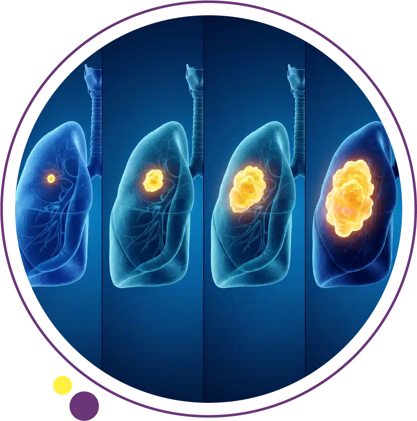
Stage I: Localized Lung Cancer
Stage I lung cancer is entirely confined to the lung with no lymph node involvement and no distant spread. Stage IA tumors are 3 cm or smaller and may be further subdivided into IA1 (1 cm or smaller), IA2 (larger than 1 cm but 2 cm or smaller), and IA3 (larger than 2 cm but 3 cm or smaller). Stage IB tumors are larger than 3 cm but not more than 4 cm and remain confined to the lung without involving major airways, the visceral pleura, or regional lymph nodes.
Stage I lung cancer represents the most favourable clinical scenario. Five-year survival rates for stage IA range from approximately 77 to 92 percent depending on the specific substage, with the smallest tumors (IA1) achieving the highest survival. Surgical resection, typically lobectomy, is the standard of care for surgically fit patients with stage I disease. Stereotactic body radiotherapy (SBRT) is a curative alternative for patients who cannot tolerate surgery due to poor lung function or other medical comorbidities. Stage I lung cancer is unfortunately uncommon at diagnosis because of the absence of early symptoms, which is precisely why CT screening in high-risk populations and prompt investigation of suspicious symptoms is so clinically valuable. For guidance on recognising those symptoms, our article on how to know if you have lung cancer provides a practical framework. The chest X-ray may reveal a lung mass, but CT is needed to characterise it fully for staging.
Stage II: Locally Contained With Limited Nodal Involvement
Stage II lung cancer involves either a larger primary tumor without lymph node involvement, or a smaller primary tumor with involvement of the ipsilateral peribronchial or hilar lymph nodes. Stage IIA describes tumors larger than 4 cm but not more than 5 cm without lymph node involvement, or tumors of 3 cm or smaller with ipsilateral hilar or peribronchial nodal involvement. Stage IIB includes tumors between 5 and 7 cm without nodal involvement, or smaller tumors with nodal involvement and specific additional features such as involvement of the main bronchus, the parietal pleura, or atelectasis of the entire lung.
Five-year survival rates for stage II lung cancer are in the range of 45 to 60 percent depending on the substage. Surgery remains the primary treatment approach for stage II disease in patients who are fit for resection, typically combined with adjuvant chemotherapy to reduce the risk of systemic recurrence. Nodal involvement identified at surgery may modify the post-operative treatment recommendation. Accurate pre-operative staging, including assessment of mediastinal lymph nodes by CT and often confirmed by EBUS (endobronchial ultrasound-guided biopsy), is essential for treatment planning. The imaging services at Images support the CT staging component of this evaluation.
Stage III: Locally Advanced Disease
Stage III lung cancer represents locally advanced disease where the tumor or its regional lymph node spread has grown beyond what is resectable with curative intent in most cases, but has not yet produced distant organ metastases. Stage III is divided into three substages that carry significantly different clinical implications and treatment approaches.
Stage IIIA includes tumors with ipsilateral mediastinal or subcarinal lymph node involvement (N2 disease) and tumors that have invaded specific adjacent structures such as the chest wall, diaphragm, or pericardium. Some IIIA cases are potentially operable, particularly when N2 disease is limited and confirmed to a single node station, and are treated with multimodality therapy combining chemotherapy, surgery, and radiation. Stage IIIB involves more extensive N2 disease or contralateral nodal involvement (N3 disease) and is generally not surgically resectable; concurrent chemoradiotherapy followed by consolidation immunotherapy is the current standard of care. Stage IIIC combines any N3 nodal involvement with a T4 tumor and is treated with the same concurrent chemoradiotherapy approach. Five-year survival rates for stage III range from approximately 10 to 36 percent depending on the substage and treatment received. Accurate determination of which nodes are involved and the resectability of the primary tumor requires high-quality CT imaging of the chest and mediastinum alongside functional assessment tools.
Stage IV: Metastatic Lung Cancer
Stage IV lung cancer is defined by the presence of distant metastatic spread beyond the chest. Stage IVA covers three specific scenarios: a separate tumor nodule in the contralateral (opposite) lung, malignant pleural or pericardial effusion, or a single distant extrathoracic organ metastasis. Stage IVB is defined by multiple extrathoracic metastases in one or more distant organs, which may include the brain, liver, adrenal glands, bones, or other sites.
Stage IV is the most commonly diagnosed stage at presentation in lung cancer, reflecting the late appearance of symptoms discussed earlier. Five-year survival rates for stage IV lung cancer have historically been below 10 percent, but the introduction of immunotherapy and molecular targeted therapies has meaningfully extended median survival for specific patient populations with responsive tumors. Patients with EGFR mutations receiving targeted oral therapy, or patients with high PD-L1 expression receiving first-line pembrolizumab, can achieve durable responses lasting years in some cases. Brain MRI is an essential component of stage IV staging for NSCLC, as intracranial metastases are common and their presence influences both systemic and local treatment decisions. The CT of chest and abdomen at Images identifies and characterises extrathoracic metastatic deposits as part of the staging workup.
Small Cell Lung Cancer Staging: Limited vs Extensive
Small cell lung cancer uses a simplified two-stage classification rather than the full TNM system used for NSCLC. Limited stage SCLC is defined as disease confined to one hemithorax and the regional mediastinal and ipsilateral supraclavicular lymph nodes, such that the entire disease volume can be encompassed within a single radiation treatment field. Extensive stage SCLC encompasses any disease beyond this definition, including malignant pleural effusion, contralateral supraclavicular involvement, and distant organ metastases.
The importance of this distinction is that limited stage SCLC is treated with concurrent chemoradiotherapy with curative intent, while extensive stage disease is treated with systemic platinum-based chemotherapy and immunotherapy with disease control and palliation as the primary goals. Approximately 60 to 70 percent of SCLC patients have extensive stage disease at diagnosis, reflecting the extremely rapid growth and early dissemination characteristic of this tumor type. Brain MRI at staging is performed in all SCLC patients regardless of stage because brain metastases are very common in this disease. The 3 Tesla brain MRI available at Images is particularly sensitive for detecting small brain metastases that can influence treatment planning. For an understanding of how SCLC differs from NSCLC in biological behavior and treatment, our article on types of lung cancer covers each subtype in detail.
How Imaging Determines Stage in Practice
The staging workup for lung cancer is an imaging-intensive process. CT of the chest and abdomen with contrast is the foundational study, defining the primary tumor’s size and local extent (T descriptor), evaluating mediastinal lymph node size for N descriptor assessment, and identifying adrenal, liver, and other abdominal metastases for M descriptor assignment. CT provides the anatomical roadmap that staging is built upon.
Brain MRI is added for all stage III and IV NSCLC and all SCLC patients to assess for intracranial disease. PET-CT, which maps metabolically active disease throughout the body, is increasingly used for NSCLC staging because it detects mediastinal and distant metastases with greater sensitivity than CT alone and can identify occult metastases that alter the stage and treatment approach. Endobronchial ultrasound (EBUS) provides minimally invasive tissue sampling of mediastinal lymph nodes to confirm or exclude N2 and N3 involvement when CT and PET findings are equivocal. Together, these imaging and tissue sampling tools build the complete staging picture. The imaging services at Images provide the CT and MRI components of this pathway for patients in Kuwait.
Frequently Asked Questions
What is the most curable stage of lung cancer?
Stage I lung cancer has the highest survival rates and the greatest probability of cure with surgery or SBRT. Stage IA1, the smallest tumor category, achieves five-year survival rates approaching 90 percent in some series. The challenge is that stage I lung cancer almost never produces symptoms and is typically found either incidentally on imaging performed for another reason or through CT lung screening in high-risk individuals. Earlier-stage discovery through proactive screening remains the most reliable route to encountering curable lung cancer.
What does stage IV lung cancer mean for treatment?
Stage IV means the cancer has spread to distant organs and curative intent surgery is no longer possible. However, treatment at stage IV is not simply palliative for all patients. Molecular targeted therapy for EGFR, ALK, ROS1, KRAS, and other actionable mutations can produce prolonged disease control. Immunotherapy achieves durable responses in patients with high PD-L1 expression. The goal shifts from cure to disease control and quality of life preservation, and modern systemic therapies have meaningfully extended survival compared to chemotherapy alone. Molecular profiling at diagnosis is essential for identifying which treatments are most likely to be effective.
Can stage III lung cancer be cured?
Some stage III lung cancer cases can be cured, particularly stage IIIA with limited N2 disease when treated at specialist multidisciplinary centers with combined modality therapy. Stage IIIB and IIIC are less frequently curable but concurrent chemoradiotherapy followed by durvalumab immunotherapy has improved outcomes significantly, with some patients achieving long-term disease-free survival. Stage III represents a heterogeneous group where individualised assessment of resectability and treatment intensity is critical.
Does chemotherapy change the stage of lung cancer?
Chemotherapy or other systemic treatments can produce radiological downstaging, meaning the tumor shrinks on imaging to the point where it appears to meet criteria for a lower stage. However, the formal stage at diagnosis remains the reference point for statistical and prognostic purposes. Radiological response is assessed at interim and end-of-treatment timepoints using CT and MRI, and significant response to neoadjuvant therapy can sometimes open a surgical window that was not available at initial presentation. CT imaging at Images supports this response monitoring throughout the treatment course.
Is brain MRI necessary for all lung cancer patients?
Brain MRI is recommended for all small cell lung cancer patients regardless of clinical symptoms because of the high frequency of asymptomatic brain metastases. For NSCLC, brain MRI is recommended for stage III and IV patients and for any patient with neurological symptoms. For stage I and II NSCLC without neurological symptoms, brain MRI is not routinely required but may be performed in specific clinical circumstances. The 3 Tesla brain MRI at Images provides the resolution needed to detect even small intracranial metastases that would be clinically significant for treatment planning.
Understanding Your Stage and What It Means Going Forward
Lung cancer staging is not a sentence; it is a starting point. Knowing the stage precisely allows the clinical team to design the most appropriate treatment strategy, set realistic goals, and monitor progress objectively. For patients with early-stage disease, accurate staging confirms that curative treatment is the right path. For patients with advanced disease, it identifies the best systemic treatment options and helps align expectations with what modern oncology can realistically achieve.
Images Diagnostic Center provides CT scanning and MRI across three Kuwait branches to support the full lung cancer staging process and ongoing treatment monitoring:
To arrange imaging as part of your lung cancer staging or monitoring, contact Images directly.