
Lung cancer is one of the most challenging cancers to detect early because in its initial stages, when treatment is most likely to be curative, it almost never produces symptoms. The lung tissue itself has no pain receptors, meaning a tumor can grow for months or years in the periphery of the lung without causing any sensation that would prompt a patient to seek medical attention. By the time symptoms do appear, the disease is frequently at a stage where options are narrower.
Knowing how to recognise the early signals, understanding which risk factors make early investigation more urgent, and being aware of the role that screening imaging plays for high-risk individuals can make a genuine and measurable difference in outcomes. In this article, we will discuss the key warning signs that may indicate lung cancer, what makes them distinctive from ordinary respiratory complaints, the risk profile that should prompt earlier and more proactive investigation, how imaging confirms or rules out lung cancer, and when to act rather than wait.
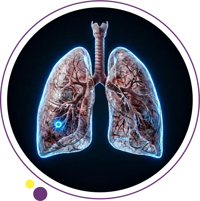
Why Lung Cancer Is So Often Found Late
The lung is anatomically large and physiologically resilient. A growing tumor in the outer zones of the lung can reach a significant size before it causes any obstruction, bleeding, or pressure on surrounding structures. Symptoms typically only develop when the tumor has grown large enough to obstruct an airway, invade the pleura or chest wall, spread to mediastinal structures, or produced distant metastases that cause symptoms in another organ entirely. This biological reality is the primary driver of the well-documented pattern of late diagnosis in lung cancer globally.
A secondary factor is symptom overlap. Cough, breathlessness, and chest discomfort are among the most common complaints in general practice and in the vast majority of cases are caused by far less serious conditions including respiratory infections, asthma, GERD, and anxiety. The challenge is identifying the minority of cases where these same symptoms represent something more serious. Understanding what distinguishes lung cancer symptoms from their benign mimics is the critical skill, and it ultimately comes down to persistence, progression, and the clinical context of the individual patient. Our comprehensive article on lung cancer covers the full picture of the disease from causes through to treatment for readers who want a complete overview.
Key Warning Signs That May Indicate Lung Cancer
A Cough That Will Not Go Away
A persistent cough lasting more than three weeks without a clear and resolving cause is one of the most significant early warning signs for lung cancer. In smokers, there is a particular risk that a change in the character, frequency, or severity of an existing chronic cough gets attributed to the smoking itself and is not investigated. A cough that has changed, a cough that has become productive of blood-tinged sputum, or a cough that is accompanied by a new wheeze or voice change should never be normalised as simply a smoker’s cough. It warrants imaging.
In non-smokers, a persistent unexplained cough carries the same investigative priority even though the prior probability of lung cancer is lower. The key question is not about the cough in isolation but about whether it is resolving as expected for an ordinary respiratory cause. If three to four weeks of appropriate management has not produced clear improvement, a chest X-ray followed by CT of the chest if the X-ray is abnormal or clinically suspicious is the standard pathway.
Coughing Up Blood
Haemoptysis, even a single episode of blood in the sputum, is a symptom that demands prompt medical evaluation without exception. While the majority of haemoptysis episodes are caused by benign conditions such as bronchitis, respiratory infection, or bronchiectasis, lung cancer is on the differential diagnosis whenever blood appears in sputum, and it cannot be excluded without imaging. A small streak of blood in sputum once in a patient with relevant risk factors should be treated with the same urgency as a larger bleed.
Imaging for haemoptysis begins with a chest X-ray and is almost always followed by CT of the chest, which provides far more detail than plain X-ray for identifying bronchial or pulmonary masses, vascular abnormalities, and other structural causes. A normal chest X-ray in the context of haemoptysis does not exclude a central airway lesion, and CT is therefore required before lung cancer can be genuinely ruled out. The CT service at Images is available across three Kuwait branches for prompt evaluation of these presentations.
Voice Changes and Hoarseness
A new or worsening hoarse voice that has persisted for more than two to three weeks without an obvious ENT cause should prompt consideration of a thoracic origin. The left recurrent laryngeal nerve loops down into the chest around the aortic arch before returning upward to supply the left vocal cord. Tumors growing in the left upper lobe, the mediastinum, or near the aortic arch can compress or invade this nerve, causing left vocal cord paralysis and a characteristic hoarse or breathy voice quality.
When a patient presents with hoarseness and laryngoscopy reveals left vocal cord paralysis without a primary ENT cause, chest imaging is mandatory to exclude a mediastinal or pulmonary mass. This symptom typically indicates a tumor that has grown beyond the lung into the mediastinum, meaning at least locally advanced disease, making prompt imaging even more important. Our detailed article on lung cancer symptoms covers this and all other warning signs in full clinical detail.
Recurrent Chest Infections in the Same Location
A pattern of recurrent pneumonia or bronchitis consistently involving the same lobe or segment of the lung is a red flag that warrants investigation for an obstructing lesion. When a central airway tumor partially or fully obstructs a bronchus, secretions accumulate in the lung distal to the obstruction, creating conditions that favour repeated bacterial infections. The patient may respond to antibiotics each time, but the infection recurs because the underlying obstructive cause has not been addressed.
Two or more pneumonia episodes in the same lung region within a twelve-month period should prompt CT imaging to evaluate for an obstructing central lesion. Chest X-ray may show the infection but may not clearly reveal a central mass, particularly if the consolidation obscures the underlying tumor. CT of the chest provides the cross-sectional detail needed to identify a bronchial lesion at the root of the obstructive pattern. This is one of several reasons why CT imaging is preferred over plain X-ray alone when lung cancer is a genuine clinical concern.
Unexplained Breathlessness
Breathlessness that has developed progressively over weeks to months without an obvious cause such as new-onset cardiac failure, significant anaemia, or a change in physical fitness deserves clinical evaluation including chest imaging. A lung tumor obstructing an airway can cause collapse of the affected lung segment, reducing effective ventilation. A pleural effusion (fluid accumulating between the lung and chest wall) secondary to pleural involvement can compress the lung and produce breathlessness that worsens gradually as the effusion grows. Both of these mechanisms are visible on imaging.
Progressive breathlessness in a patient over fifty with a significant smoking history should be treated as potentially serious until proven otherwise, even if an alternative explanation such as COPD seems likely. The possibility that a new central or obstructive mass is contributing to worsening breathlessness on a background of existing lung disease is real and should not be dismissed without imaging. The chest X-ray is the practical first step, and CT follows when the X-ray raises a concern or when clinical suspicion remains despite a normal or near-normal X-ray. At Images Diagnostic Center, both imaging studies are available promptly.
Unexplained Weight Loss and Fatigue
Significant unintentional weight loss of more than five percent of body weight over six months, accompanied by persistent fatigue, in a patient with lung cancer risk factors is a combination that warrants a full clinical investigation including chest imaging. These systemic symptoms reflect the metabolic burden of a growing tumor and the body’s inflammatory response to it. They are more prominent in advanced disease but can occasionally appear in locally advanced lung cancer before extensive metastatic spread has occurred.
When weight loss and fatigue are the presenting complaints and the cause is not immediately apparent from initial assessment, imaging of the chest is typically part of the investigative workup. The CT scan provides a simultaneous view of the chest, mediastinum, and upper abdomen, meaning a single study can evaluate both the chest and the liver and adrenal glands for evidence of intrathoracic or extrathoracic malignancy. For an overview of all the warning signs and how they relate to each other and to specific lung cancer types, our article on types of lung cancer provides useful complementary context.
Bone Pain or Neurological Symptoms
When lung cancer has spread to distant organs, it produces symptoms in those organs. Persistent bone pain, particularly in the back, hips, or ribs, without a traumatic explanation may reflect skeletal metastases from a lung primary. New neurological symptoms including severe headache, visual disturbance, speech difficulty, seizures, or limb weakness in a patient with respiratory symptoms or risk factors may indicate brain metastases, which are common in both non-small cell and small cell lung cancer.
In these presentations, the neurological or bone symptoms may actually be the first thing that brings the patient to medical attention, and the lung primary is then identified when imaging is performed. Brain MRI is the most sensitive study for intracranial metastases, and 3 Tesla MRI at Images provides the resolution needed to detect even small cortical or subcortical lesions. CT of the chest is then performed to identify the primary lung tumor. When symptoms outside the lung are present alongside any respiratory symptoms, acting promptly on both is essential.
Who Is at Highest Risk and Why Risk Matters for Early Detection
The clinical significance of any of the above symptoms is substantially influenced by the patient’s risk profile. The highest-risk individuals are current or former heavy smokers aged fifty and above who have smoked for twenty or more pack-years (a pack-year is one pack of cigarettes per day for one year). In this population, the prior probability of lung cancer as the cause of a persistent respiratory symptom is meaningfully higher than in a young, never-smoking adult, which justifies a lower threshold for investigation and a faster escalation to CT imaging.
Other risk factors that elevate the clinical index of suspicion include occupational exposure to asbestos, arsenic, chromium, or diesel exhaust; a family history of lung cancer; a personal history of prior lung cancer or prior head and neck cancer treated with radiation; and known chronic lung diseases such as pulmonary fibrosis that are independently associated with elevated lung cancer risk. Non-smokers who develop respiratory symptoms in the presence of other risk factors should not be falsely reassured by the absence of a smoking history. Lung cancer in never-smokers is a genuine and growing clinical entity. The full imaging services at Images are available to support evaluation in all of these risk contexts.
The Role of Low-Dose CT Lung Screening
For individuals in the highest-risk group, particularly current and former heavy smokers aged fifty to seventy-seven, annual low-dose CT lung screening has been demonstrated in large clinical trials to reduce lung cancer mortality by detecting cancers at earlier and more treatable stages before symptoms develop. The National Lung Screening Trial (NLST) in the United States showed a 20 percent reduction in lung cancer mortality with annual low-dose CT compared to chest X-ray in this population, and subsequent trials have confirmed these findings with even greater mortality reductions in some analyses.
Low-dose CT screening uses a significantly reduced radiation dose compared to a standard diagnostic CT while still producing images of sufficient quality to detect lung nodules as small as a few millimetres. Identified nodules are then followed according to established protocols to determine which require further investigation and which can be monitored with surveillance imaging. For patients in Kuwait who qualify based on their smoking history and age, speaking with their physician about whether CT lung screening is appropriate is a genuinely important conversation. CT scanning at Images supports both diagnostic evaluation and physician-directed lung assessment.
When to Act: A Clear Decision Framework
The decision to seek medical evaluation and imaging does not require certainty that lung cancer is present. It requires recognition that a symptom pattern is persistent, progressive, or occurring in the context of a risk profile that justifies investigation. As a general guide: any respiratory symptom that has not resolved within three to four weeks and has no clear benign explanation should prompt a consultation. Any haemoptysis, regardless of amount, should prompt same-week evaluation. Any new neurological symptom in a patient with respiratory symptoms or risk factors should prompt urgent imaging. Any systemic symptoms including unexplained weight loss, bone pain, or fatigue in a patient over fifty with smoking history should be investigated without delay.
The imaging pathway typically begins with a chest X-ray but should escalate quickly to CT chest when clinical suspicion is present, because CT detects far more lesions and characterises them far better than plain X-ray. A normal chest X-ray in a clinically concerning presentation does not exclude lung cancer and should not be used as reassurance alone. Our article on CT scan uses explains in detail why CT is the modality of choice for thoracic evaluation when a significant lung pathology is being considered. At Images, CT and chest X-ray are available at all three Kuwait branches for prompt evaluation.
Frequently Asked Questions
Can lung cancer be detected without symptoms?
Yes. Low-dose CT lung screening detects early-stage lung cancer in high-risk individuals before any symptoms develop, and this is precisely why screening improves outcomes. Additionally, lung nodules are sometimes found incidentally on CT scans performed for other clinical reasons. Early-stage lung cancer found incidentally or through screening has a substantially better prognosis than symptomatic lung cancer diagnosed after clinical presentation, underscoring the value of proactive imaging in high-risk individuals.
Is a chest X-ray enough to check for lung cancer?
A chest X-ray is a useful first step but it has significant limitations for lung cancer detection. Small peripheral tumors, central airway masses, and lesions in anatomically challenging areas can be invisible or very subtle on plain X-ray. Clinical trials comparing chest X-ray to low-dose CT for lung cancer screening showed no mortality benefit from X-ray screening, whereas CT screening produced clear benefit. When lung cancer is a genuine clinical concern, CT of the chest is the appropriate investigation rather than chest X-ray alone.
What is the most common early sign of lung cancer?
A persistent cough that does not resolve over three to four weeks is the most commonly reported early symptom, though strictly speaking it reflects locally advanced rather than truly early disease. Truly early lung cancer (stage I) almost never produces symptoms. This is the fundamental challenge of lung cancer detection and the reason why screening high-risk individuals with CT before symptoms develop is the most effective strategy for identifying disease at a genuinely early and curable stage.
Can non-smokers get lung cancer?
Yes. Approximately 10 to 15 percent of lung cancer cases globally occur in people who have never smoked. Non-smoker lung cancer is more commonly of the adenocarcinoma subtype and is frequently associated with targetable molecular mutations including EGFR and ALK alterations. Risk factors for non-smoker lung cancer include radon exposure, secondhand smoke exposure over many years, air pollution, occupational carcinogens, and genetic susceptibility. Non-smokers should not be falsely reassured by their non-smoking status when persistent respiratory symptoms are present.
How quickly should I see a doctor if I notice these symptoms?
Any haemoptysis or sudden severe neurological symptom warrants urgent same-day or same-week evaluation. Persistent cough, unexplained breathlessness, or unexplained weight loss lasting more than three to four weeks without resolution should prompt a medical consultation within days rather than weeks, particularly if any lung cancer risk factors are present. Early investigation does not guarantee early cancer detection, but waiting always reduces the probability that any cancer present is found at a stage where curative treatment is possible. Arranging a CT scan at Images in Kuwait can be done promptly once your doctor has made a referral.
Early Action Is the Most Powerful Tool Available
Knowing how to recognise the warning signs of lung cancer, understanding that a normal chest X-ray is not sufficient reassurance when clinical suspicion is present, and appreciating the value of screening in high-risk individuals are the three most practically important things anyone can take from this article. Lung cancer detected before it spreads carries a fundamentally different prognosis from the same disease found after metastasis has occurred. The window for curative treatment is real, but it requires acting on symptoms rather than waiting for them to become severe.
Images Diagnostic Center provides chest X-ray, CT scanning, and MRI across three Kuwait branches to support the full diagnostic evaluation of suspected lung cancer:
To arrange a chest CT or discuss the right imaging pathway for your situation, contact Images directly.