
X-ray is one of the oldest and most widely used imaging tools in medicine, yet its range of clinical applications is broader than many people realize. Most patients associate X-ray with broken bones, but the reality is that it plays a meaningful diagnostic role across many areas of the body and serves as the first step in clinical pathways for a wide variety of medical conditions.
Understanding the specific uses of X-ray imaging helps patients appreciate why their doctor has requested one, what it will and will not show, and when a different type of imaging might be more appropriate. In this article, we will discuss the main medical uses of X-ray, which body systems it evaluates most effectively, how it compares to other imaging modalities in specific contexts, and when it remains the most clinically sensible first step in a diagnostic workup.
How X-Ray Produces Diagnostic Images
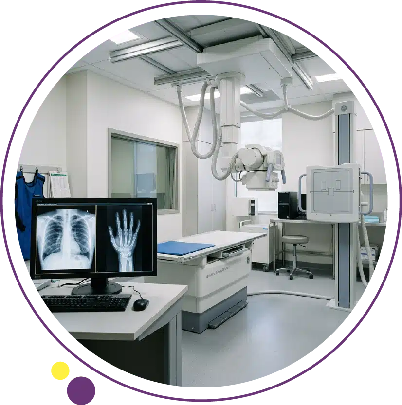
X-ray imaging creates images by passing a controlled beam of ionizing radiation through the body. Dense tissues like bone absorb most of the beam and appear white on the resulting image, while softer tissues allow more of the beam to pass through and appear in varying shades of grey. Air-filled spaces such as the lungs appear dark because they absorb almost none of the beam. This natural contrast between different tissue types is what gives X-ray its diagnostic value.
Modern digital X-ray systems produce images electronically rather than on film, making them faster to acquire, easier to enhance and transmit, and lower in radiation dose than older equipment. Digital detectors capture a wide dynamic range of information in a single exposure, and radiologists can adjust image contrast and brightness during review to optimize visualization of different tissue types in the same image. This technical improvement has made digital X-ray both more informative and more practical than its predecessors across all of its established clinical uses.
Bone and Musculoskeletal Uses
Fracture Detection and Classification
The detection and characterization of bone fractures is the most well-known use of X-ray imaging, and it remains the standard first-line approach for this purpose in both emergency and outpatient settings. X-ray clearly shows cortical disruptions, displacement, angulation, comminution, and alignment in most types of fractures. For the majority of fractures encountered in clinical practice, an X-ray provides all the information needed to plan conservative or surgical management.
X-ray for fracture assessment is performed using at least two views taken at perpendicular angles to provide a three-dimensional understanding of the injury. In some complex fractures, particularly around joints or in the spine, follow-up with CT scan is requested to better assess fracture geometry before surgical planning. But in the majority of cases, the initial X-ray is both the starting point and the clinically sufficient study for fracture management decisions.
Joint Disease and Arthritis
X-ray is the primary imaging tool for assessing osteoarthritis, rheumatoid arthritis, and other joint conditions. It clearly shows joint space narrowing, which reflects cartilage loss, as well as osteophyte formation, subchondral sclerosis, erosions, and bone deformity. These findings are used to grade the severity of arthritis, monitor its progression over time, and guide decisions about conservative management or joint replacement surgery.
In rheumatoid arthritis, serial X-rays of the hands and feet are a standard part of disease monitoring because they track erosive changes and the effectiveness of treatment in slowing structural damage. X-ray provides this longitudinal picture in a practical and cost-effective way that other imaging modalities cannot replicate with the same efficiency. For soft tissue changes within joints, including synovitis, cartilage detail, and tendon assessment, MRI or ultrasound is more informative, and the two approaches are often used as complementary rather than competing tools in rheumatological care.
Scoliosis and Spinal Alignment
Full-length standing X-rays of the spine are the standard method for diagnosing and monitoring scoliosis, measuring the degree of spinal curvature, and assessing treatment response in children and adults. The ability to image the full spine in a weight-bearing position, capturing functional alignment rather than supine positioning, makes X-ray uniquely suited to scoliosis assessment in a way that MRI or CT cannot replicate practically in routine monitoring.
Spinal X-rays are also used to assess vertebral alignment after trauma, evaluate degenerative disc disease with reduced disc height, identify spondylolysis, and monitor patients with instrumented spinal fusions post-operatively. While MRI provides far more detail about disc, cord, and nerve root pathology, plain X-ray of the spine retains an important and irreplaceable role in the structural and alignment assessment of spinal conditions. For patients in Kuwait managing spinal conditions, digital X-ray at Images provides the imaging backbone for many of these ongoing monitoring needs.
Chest and Respiratory Uses
Lung and Respiratory Conditions
The chest X-ray is one of the most performed radiological studies in clinical medicine globally and is the front-line investigation for a wide range of respiratory and cardiac presentations. It provides an immediate overview of the lungs, airways, pleural spaces, mediastinum, heart, and ribs in a single image that can be acquired in less than a minute.
Conditions commonly assessed and identified on chest X-ray include pneumonia, pleural effusion, pneumothorax, atelectasis, pulmonary edema, rib fractures, and mediastinal widening. A chest X-ray is typically the first imaging study ordered for patients presenting with cough, fever, chest pain, shortness of breath, or hemoptysis in both primary care and emergency settings. The speed and simplicity of the study make it practical in almost any clinical environment. Patients with findings suggesting a lung mass or other detailed pulmonary pathology are subsequently referred for a CT scan of the chest, which provides cross-sectional detail that X-ray cannot offer.
Cardiac Assessment on Chest X-Ray
While cardiac imaging with dedicated modalities such as echocardiography, Cardiac MRI, or Cardiac CT provides far more functional and structural detail, a chest X-ray can identify cardiac enlargement, pulmonary vascular congestion, and mediastinal abnormalities that point toward cardiac pathology. Cardiomegaly visible on chest X-ray, for example, is a prompt to arrange further cardiac evaluation. This makes the chest X-ray a useful first screening tool in the cardiac assessment pathway, even if it does not replace dedicated cardiac imaging for definitive assessment.
Preoperative and Health Screening Assessments
Chest X-ray is routinely included in preoperative assessment protocols for patients undergoing major surgery, particularly in older adults and those with known or suspected cardiorespiratory conditions. It provides a baseline assessment of lung and cardiac status that helps anesthetists and surgeons plan the procedure safely. In health screening programs, chest X-ray is often used as an initial study to assess lung and cardiac health, with follow-up imaging arranged for patients whose results suggest further investigation is warranted. The full range of imaging services at Images supports the comprehensive diagnostic pathway from initial X-ray through to advanced cross-sectional and specialized imaging as clinical needs evolve.
Abdominal Uses of X-Ray
Bowel Obstruction and Perforation
Abdominal X-ray is used in specific emergency scenarios where quick assessment of bowel gas distribution, obstruction, or free air is needed. Dilated bowel loops suggesting obstruction, free intraperitoneal air indicating perforation, and abnormal gas patterns can all be identified on a plain abdominal X-ray. While CT has largely replaced plain X-ray for abdominal emergencies in settings where CT is readily available, abdominal X-ray retains practical utility in rapid triage situations and in settings where immediate CT access is limited.
Plain abdominal X-ray is also used to identify radiopaque foreign bodies that have been ingested or to confirm the position of indwelling devices such as nasogastric tubes or urinary catheters when other methods are not immediately available. For detailed abdominal organ assessment including the liver, kidneys, and bowel wall, ultrasound or CT is far more informative and is the preferred modality in non-emergency abdominal imaging. Our published guide on CT scan uses explains where CT adds clinical depth beyond what plain X-ray can show in abdominal and other diagnostic contexts.
Kidney Stones and Calcifications
Calcified kidney stones, gallstones (when calcified), and vascular calcifications can sometimes be identified on plain abdominal X-ray. However, a significant proportion of urinary tract stones are not radiopaque and will not appear on a standard X-ray, making ultrasound or CT the more reliable imaging options for suspected renal colic or urinary tract stone disease. When calcifications are identified on an abdominal X-ray, they may prompt further targeted imaging to characterize their nature and clinical significance more precisely. The ultrasound service at Images is well suited to this follow-up role, offering real-time soft tissue assessment of the kidneys and urinary tract alongside the stone detection sensitivity that plain X-ray cannot match.
Dental and Skull Uses
Dental X-rays are a fundamental tool in dentistry, providing detailed images of tooth structure, root morphology, inter-dental bone levels, and the position of unerupted or impacted teeth. Different types of dental X-ray are used for different purposes: periapical views show individual tooth roots and surrounding bone, bite-wing views assess inter-proximal bone loss and early caries between teeth, and panoramic radiographs provide a full overview of the dentition and jaw in a single wide-field image.
Skull X-rays have become less commonly used in clinical practice following the wider availability of CT, which provides far more detail for skull and intracranial assessment. However, skull X-ray is still used in some settings for assessing specific bone pathology, identifying foreign bodies in the orbital region, and in forensic or resource-limited contexts. For patients with head trauma or suspected intracranial pathology, CT is the appropriate study. At Images Diagnostic Center, patients can access digital X-ray, CT, and MRI services, allowing the appropriate imaging modality to be selected based on the specific clinical question being addressed at each stage of investigation.
Bone Density Assessment and X-Ray
While standard X-ray can suggest reduced bone density in advanced cases, it is not a reliable or sensitive tool for diagnosing osteoporosis. Bone density changes visible on plain X-ray typically reflect losses of 30 percent or more, meaning significant disease may already be present before it becomes apparent on a standard X-ray. The dedicated tool for bone mineral density measurement is a DEXA scan, which uses a very low-dose X-ray technique specifically calibrated to measure bone density accurately and compare it against normative reference values.
Plain X-rays of the spine or hip may incidentally suggest osteoporosis through characteristic findings such as vertebral compression fractures or cortical thinning, and these findings should prompt formal DEXA scanning rather than relying on the X-ray appearance alone for diagnosis. For patients in Kuwait who have been identified as having osteoporosis risk factors or who have had an incidental finding on a spinal X-ray, the DEXA scan service at Images provides the accurate and dedicated bone density measurement that clinical guidelines recommend for formal diagnosis and treatment planning.
X-Ray in Pediatric Medicine
X-ray is widely used in pediatric care for the same range of indications as in adults, but with an additional emphasis on minimizing radiation exposure given the longer lifetime over which any cumulative risk would accumulate. Modern digital X-ray equipment operates at significantly lower doses than older systems, and pediatric protocols are specifically optimized to reduce exposure while maintaining diagnostic image quality. Pediatric radiographers are trained to apply the ALARA principle, ensuring radiation is kept as low as reasonably achievable for each individual study.
Common pediatric uses include assessment of respiratory infections such as childhood pneumonia on chest X-ray, fracture evaluation in children following falls or trauma, assessment of bone age in growth and developmental concerns, and swallowed or inhaled foreign body localization. For soft tissue and joint assessment in children where radiation avoidance is particularly important, ultrasound is often preferred as an alternative. The Ultrasound and Doppler service at Images provides a radiation-free option that is well suited to pediatric soft tissue and joint assessment when X-ray is not appropriate or sufficient.
X-Ray as Part of a Broader Diagnostic Pathway
One of the most important things to understand about X-ray is that it rarely stands alone as the complete answer to a complex diagnostic question. In the majority of clinical pathways, X-ray serves as the fast, accessible, and practical first step that establishes a baseline, identifies obvious pathology, and directs the next stage of investigation when more detail is needed.
For example, a patient presenting with back pain may first have a lumbar spine X-ray to assess bony alignment, disc space height, and degenerative changes. If the clinical picture suggests nerve root compression, disc herniation, or spinal cord involvement, an MRI of the lumbar spine is the next step, providing the soft tissue detail that X-ray cannot offer. Similarly, a chest X-ray showing a possible pulmonary mass will be followed by a CT chest for detailed characterization before any further clinical decision is made. This layered approach to imaging, starting with the most accessible and informative first-line study and escalating as needed, is both clinically sound and practically efficient. Patients exploring all the diagnostic imaging options available to them in Kuwait can review the full services at Images to understand how each modality fits into this broader picture.
Frequently Asked Questions
What conditions can an X-ray diagnose?
X-ray is used to diagnose and assess a wide range of conditions including fractures, dislocations, joint disease, osteoarthritis, scoliosis, pneumonia, pneumothorax, pleural effusion, cardiomegaly, bowel obstruction, and certain foreign bodies. It provides immediate information about bony structures and air-filled spaces, and can indicate when further imaging with CT, MRI, or ultrasound is warranted for more detailed assessment of soft tissues or internal organ pathology.
Is X-ray better than CT scan for fractures?
For most straightforward fractures, plain X-ray is both sufficient and more practical than CT as an initial study. CT is reserved for complex fractures, particularly around joints or in the spine, where three-dimensional detail is needed for surgical planning. Starting with X-ray and escalating to CT when additional detail is clinically required is the standard and evidence-based approach in most musculoskeletal trauma scenarios.
Can X-ray show soft tissue injuries like ligament tears?
No. X-ray cannot directly visualize soft tissue structures such as ligaments, tendons, or cartilage. It can suggest soft tissue injury indirectly through findings like joint instability or unusual bony alignment, but for confirmed soft tissue diagnosis, MRI or in some cases ultrasound is required. When a patient has both bony and soft tissue injury, X-ray is typically performed first followed by MRI if soft tissue detail is clinically needed.
How many X-ray views are usually taken for a given body part?
Most body parts are imaged in at least two views taken from perpendicular angles, typically frontal and lateral. This gives a three-dimensional perspective on a two-dimensional medium and significantly improves diagnostic accuracy compared to a single view. For complex joints or specific clinical indications, additional oblique views may be requested. Your radiographer will ensure the right number and type of views are obtained for the body area being examined.
Can X-ray be done at home in Kuwait?
Yes. The Images GO home radiology service brings digital X-ray and other imaging services directly to patients who are unable to travel to a clinic. This service is particularly valuable for elderly patients, post-surgical patients, and those with mobility limitations. Home X-ray is performed using the same digital equipment standards as in-branch imaging, ensuring diagnostic image quality is maintained.
When is an X-ray not enough and a CT or MRI is needed?
X-ray is not enough when soft tissue detail is needed, when complex fracture geometry requires three-dimensional characterization, when internal organ pathology is being investigated, or when neurological conditions are suspected. In these situations, CT or MRI provides the additional diagnostic depth that the clinical question requires. Your referring doctor will determine which follow-up imaging is appropriate, and the Images team can arrange the relevant scan at the most convenient branch for you.
Arranging Your X-Ray in Kuwait
X-ray is the frontline diagnostic tool for a wide range of clinical presentations precisely because it delivers fast, reliable, and clinically relevant information in situations where quick answers matter. Understanding its specific uses across different body systems helps patients approach their imaging pathway with confidence, knowing what the X-ray will show, why their doctor requested it, and what might follow if additional detail is needed.
At Images Diagnostic Center, digital X-ray is available at all three Kuwait branches alongside the full range of advanced imaging services. If your X-ray results indicate that follow-up CT, MRI, ultrasound, or another modality is needed, all of these options are accessible within the same provider, making the transition between imaging stages straightforward and efficient.
To book a digital X-ray or ask about the right imaging option for your condition, contact the Images team directly.