
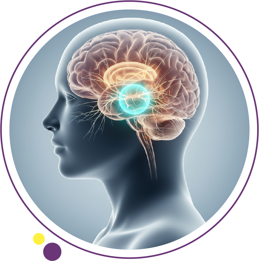
Understanding what is brain cancer begins with recognizing that it refers to the uncontrolled growth of abnormal cells within brain tissue or its surrounding structures. These tumors may arise from brain cells themselves or spread from cancers elsewhere in the body. Accurate evaluation requires advanced neuroimaging, clinical assessment, and structured radiology reporting to guide treatment decisions.
At Images Diagnostic Center in Kuwait, patients have access to comprehensive neuroimaging services including MRI scan in Kuwait, 3 Tesla MRI Kuwait, CT scan in Kuwait, and advanced contrast-enhanced imaging protocols designed to support accurate neurological diagnosis.
What is brain cancer?
one of the most dangerous types of cancer, Brain cancer is a broad term describing malignant tumors that develop within the brain or metastasize to it. Clinically, tumors are categorized as Primary brain tumors which originate in the brain, such as gliomas or meningiomas, and Secondary brain tumors, also called brain metastases, originating from cancers like lung or breast cancer.
Tumors are further classified by cell type, growth rate, World Health Organization grading, and location within the brain. The biological behavior of a tumor determines imaging strategy and urgency of intervention.
How does brain cancer develop?
Brain tumors arise when genetic mutations disrupt normal cellular regulation. These mutations may be spontaneous or influenced by prior radiation exposure, genetic syndromes, immune suppression, or metastatic spread from systemic cancer.
Unlike many other cancers, lifestyle factors have limited proven association with primary brain tumors. Imaging plays a critical role in detecting early structural changes before neurological damage becomes irreversible.
What are the most common symptoms of brain cancer?
Symptoms vary depending on tumor size, growth rate, and location. Increased intracranial pressure or focal neurological disruption typically causes clinical signs. Common warning symptoms include persistent or progressively worsening headaches, new-onset seizures in adults, vision disturbances, weakness on one side of the body, speech difficulties, memory changes, or balance problems.
Headaches related to brain tumors often worsen in the morning, intensify with coughing or straining, and do not respond to usual pain medication. Any new neurological deficit warrants urgent neuroimaging evaluation.
When should headaches or seizures prompt urgent imaging?
Immediate imaging is medically necessary when a seizure occurs for the first time in adulthood, or when headaches are associated with vomiting or altered consciousness. There is focal weakness or sensory loss, or symptoms are rapidly progressive.
In such cases, a CT scan in Kuwait may be performed initially to rule out bleeding, followed by an urgent MRI scan in Kuwait for detailed brain assessment.
Which imaging test is best for diagnosing brain cancer?
Magnetic resonance imaging as the gold standard
Magnetic resonance imaging is the most accurate modality for evaluating suspected brain tumors. An MRI scan in Kuwait provides superior soft tissue contrast, multiplanar imaging capability, detection of small lesions, evaluation of surrounding edema, and assessment of mass effect.
What makes 3 Tesla MRI more accurate for diagnosis?
A 3 Tesla MRI Kuwait system uses a stronger magnetic field compared to 1.5T MRI. This results in a higher signal-to-noise ratio, improved spatial resolution, better visualization of tumor margins, enhanced detection of small metastases, and more detailed contrast enhancement patterns.
For brain cancer evaluation, this level of detail improves diagnostic confidence and surgical planning.
How does a CT scan help in brain tumor evaluation?
A CT scan in Kuwait is often the first-line imaging tool in emergency settings. Computed tomography is particularly useful for detecting acute bleeding, identifying calcifications, evaluating bone involvement, and rapid assessment in unstable patients.
Although CT provides less soft tissue detail than MRI, it remains essential in acute neurological presentations.
What happens during a brain MRI scan?
Step-by-step patient journey
1. Arrival and screening: Completion of safety questionnaire, metal screening, review of medical history, and pregnancy status confirmation.
2. Preparation: Removal of metallic objects and IV line placement if contrast is required.
3. Scanning process: Patient lies on a padded table, head coil positioned for signal reception, and loud sounds occur during image acquisition. Scan duration typically 20 to 45 minutes.
4. After the scan: Observation if contrast was administered and resume normal activities unless advised otherwise. Motion control is critical. Even minimal head movement can degrade image quality and reduce diagnostic accuracy.
Is contrast media necessary for brain tumor detection?
Contrast-enhanced MRI uses gadolinium-based agents to highlight areas of blood-brain barrier disruption. Contrast is typically required when evaluating suspected tumors, assessing tumor grade, monitoring treatment response, or differentiating tumor from infection.
Safety considerations
Renal function is assessed when indicated, allergic history is reviewed, and hydration is recommended post-contrast. At Images Diagnostic Center, standardized contrast protocols are followed to maintain patient safety and diagnostic precision.
How fast can a CT scan report be ready?
Radiology report turnaround time depends on clinical urgency and case complexity. In urgent neurological cases, preliminary assessment may be expedited and critical findings are communicated immediately.
In routine evaluations, structured reporting ensures detailed interpretation and correlation with clinical history enhances accuracy. An accurate diagnosis imaging center Kuwait emphasizes protocol-driven imaging and subspecialty review to maintain high diagnostic standards.
What affects the accuracy of brain tumor imaging?
Several factors influence diagnostic confidence: magnet strength such as 3 Tesla MRI Kuwait, optimized imaging protocols, contrast administration timing, patient cooperation, and radiologist expertise in neuroimaging.
Adherence to internationally recognized standards, including ACR recommendations and RSNA patient safety guidance, supports consistent quality outcomes.
MRI vs CT: how do doctors choose?
Imaging selection depends on clinical context.
| Clinical Scenario | Preferred Modality |
| Acute trauma | CT scan |
| Suspected tumor | MRI scan |
| Follow-up monitoring | MRI |
| Acute hemorrhage | CT |
| Surgical planning | 3 Tesla MRI |
MRI is superior for soft tissue characterization. CT is faster and more accessible in emergencies.
Are other imaging tests used in brain cancer assessment?
In certain cases, additional studies may include MR spectroscopy, perfusion imaging, functional MRI, or CT chest or abdomen to search for primary cancer.
These techniques help differentiate tumor types and evaluate metastatic disease.
What are treatment planning considerations after imaging?
Imaging defines tumor size, location relative to eloquent brain areas, presence of edema, and mass effect or midline shift. This information guides surgical feasibility, biopsy planning, radiation targeting, and chemotherapy protocols.
Imaging follow-up is critical for monitoring recurrence or treatment response.
Post-imaging care and follow-up
After contrast MRI, drink fluids and monitor for rare allergic reactions. If biopsy is performed, follow wound care instructions and observe for neurological changes.
Patients are typically referred to neurosurgery or oncology based on imaging findings.
FAQs
- Can brain cancer be detected without symptoms?
Yes. Incidental findings occasionally appear during imaging performed for unrelated reasons. Early detection improves management planning.
- Are all brain tumors cancerous?
No. Some are benign but may still require treatment due to pressure effects on brain tissue.
- Is MRI safe?
MRI does not use ionizing radiation. Safety screening is essential for patients with implants or metallic devices.
- How long does a brain MRI take?
Typically 20 to 45 minutes depending on protocol and contrast use.
- Is radiation exposure a concern with CT scans?
CT uses ionizing radiation. Dose optimization protocols minimize exposure according to established radiology safety standards.
- Can children undergo brain MRI?
Yes. Pediatric protocols are adapted to ensure safety and minimize motion.
Making the right imaging decision
Selecting the appropriate imaging modality for suspected brain cancer depends on clinical presentation, urgency, and diagnostic goals. MRI remains the primary tool for tumor characterization, while CT plays a vital role in emergency assessment. Preparation protocols, contrast screening, standardized acquisition techniques, and structured reporting directly impact diagnostic confidence. A careful balance between image quality, patient safety, and timely interpretation ensures reliable outcomes.
Images Diagnostic Center branches in Kuwait:
Patients seeking evaluation for neurological symptoms may contact the center for further information or to schedule an appointment. The facility maintains high clinical standards in diagnostic imaging across Kuwait, emphasizing safety screening, protocol accuracy, and trusted diagnostic care to support informed medical decisions.