
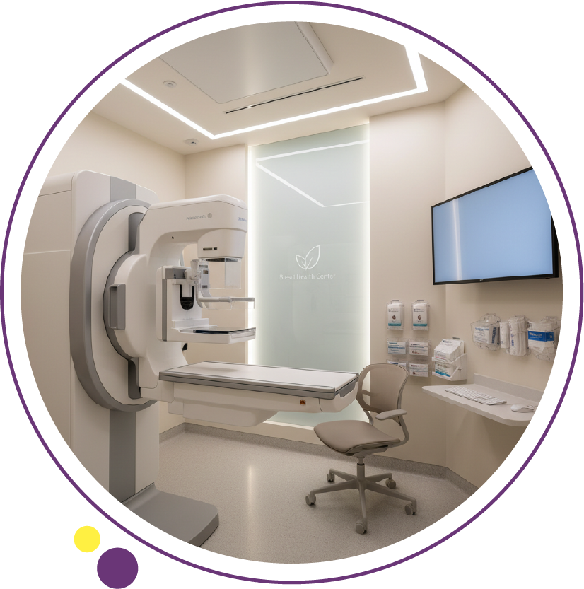
A mammogram is a specialized, low- dose X-ray examination of the breast designed to detect early signs of breast cancer and other breast abnormalities before they become clinically apparent. At Images Diagnostic Center in Kuwait, mammography is performed using standardized imaging protocols aligned with international radiology guidelines to ensure high diagnostic accuracy, radiation safety, and structured reporting.
Understanding what a mammogram is, when it is recommended, and how results are interpreted helps patients make informed decisions about breast imaging in Kuwait and ongoing breast health surveillance.
To know more about the cost of a mammogram in Kuwait you can read the guide
What is a mammogram and how does it work?
A mammogram is a type of breast imaging that uses low-dose ionizing radiation to create detailed images of breast tissue. Unlike general X-ray, mammography equipment is specifically calibrated for breast tissue contrast and spatial resolution.
During the exam:
- The breast is positioned on a flat detector plate.
- A compression paddle gently presses the breast to spread tissue evenly.
- Images are acquired in at least two standard views per breast:
- Craniocaudal view
- Mediolateral oblique view
Compression is essential because it reduces motion artifacts, improves tissue visualization, lowers radiation dose, and enhances the detection of small calcifications and masses.
The goal is to identify subtle changes such as:
- Microcalcifications
- Architectural distortion
- Asymmetries
- Solid or cystic masses
Mammography remains the primary screening tool for early detection of breast cancer, as supported by international guidance including the American College of Radiology.
Who should undergo breast screening in Kuwait?
Breast screening recommendations depend on age, risk profile, and family history. General screening guidance includes:
- Women aged 40 and above: Annual or biennial mammogram Kuwait screening depending on risk and physician advice.
- Women with family history of breast cancer: Earlier screening may be recommended.
- Known genetic predisposition: May require combined mammography and MRI scan in Kuwait.
- Women with prior breast cancer: Ongoing surveillance imaging.
Diagnostic mammography, as opposed to screening mammography, is recommended when a lump is felt or nipple discharge is present.
High-risk individuals may also benefit from adjunct imaging such as breast ultrasound or magnetic resonance imaging, depending on breast density and clinical findings.
How does a mammogram feel and how should patients prepare?
Most patients experience temporary pressure during compression. Discomfort varies but typically lasts only a few seconds per image.
Preparation checklist:
- Avoid deodorant, powder, or lotions on the chest or underarms.
- Inform staff about:
- Pregnancy possibility
- Breast implants
- Prior breast surgeries
- Previous imaging studies
- Schedule after the menstrual period if breasts are sensitive.
At Images Diagnostic Center, a structured intake process includes risk assessment and prior imaging comparison, which improves interpretation accuracy.
What do mammogram results mean?
Mammogram results are interpreted using the BI-RADS classification system, a standardized reporting framework developed by the American College of Radiology.
BI-RADS categories include:
- 0: Incomplete, additional imaging required
- 1: Negative
- 2: Benign findings
- 3: Probably benign, short-term follow-up recommended
- 4: Suspicious abnormality
- 5: Highly suggestive of malignancy
- 6: Known biopsy-proven malignancy
Structured reporting improves communication between radiologists and referring physicians and supports consistent management pathways.
When is additional imaging needed after a mammogram?
Not all abnormal mammograms indicate cancer. Additional imaging may include:
- Ultrasound in Kuwait for cyst evaluation.
- Doppler ultrasound for vascular assessment of suspicious masses.
- MRI scan in Kuwait for high-risk screening or implant evaluation.
- Image-guided biopsy for tissue diagnosis.
Dense breast tissue may reduce mammographic sensitivity. In such cases, supplementary imaging improves detection rates.
What makes breast imaging more accurate?
Diagnostic confidence in accurate diagnosis imaging center settings depends on several factors:
1. Image quality
- Proper compression
- Correct positioning
- Motion control
2. Equipment calibration
- Regular quality assurance
- Radiation dose optimization
3. Radiologist expertise
- Subspecialty training in breast imaging
- Familiarity with BI-RADS criteria
4. Comparison with prior studies
- Identifying interval change is critical.
Standardized protocols and quality control processes significantly impact early detection rates.
How fast can a mammogram report be ready?
Report turnaround time depends on:
- Screening vs diagnostic exam
- Need for additional imaging
- Complexity of findings
At structured radiology centers in Kuwait, workflow typically includes image acquisition, radiologist interpretation, and physician communication. Timely reporting supports early clinical decision-making and reduces patient anxiety.
How does mammography compare with other imaging tests?
Each modality has specific clinical roles.
Mammography vs Ultrasound
- Mammography detects microcalcifications better.
- Ultrasound differentiates cystic vs solid masses.
- Doppler ultrasound evaluates vascularity.
Mammography vs MRI
- MRI has higher sensitivity in dense breasts.
- MRI uses magnetic resonance imaging rather than radiation.
- MRI is often reserved for high-risk cases.
Mammography vs CT scan in Kuwait
- CT is not a routine breast screening tool.
- CT scan in Kuwait is used for cancer staging or systemic evaluation, not primary screening.
Mammography vs DEXA
- Bone density test (DEXA) Kuwait assesses bone mineral density.
- DEXA does not evaluate breast tissue.
Radiation safety in mammography
Mammography uses low-dose radiation. Dose optimization follows international safety principles:
- Justification of exam
- Dose minimization
- Quality control checks
The radiation exposure from a mammogram is minimal compared to diagnostic CT scans and remains within accepted safety thresholds.
Patients should inform the radiology team if pregnancy is suspected, although routine screening mammography is generally avoided during pregnancy unless clinically necessary.
Step-by-step patient journey at Images Diagnostic Center
- Appointment booking and clinical questionnaire
- Arrival and identity verification
- Breast history and risk assessment
- Imaging acquisition with certified technologist
- Image review by radiologist
- Structured report generation
- Communication of results
If further imaging or biopsy is needed, a clear follow-up plan is provided.
Contrast media in breast imaging
Routine mammography does not use contrast. Contrast may be used in:
- Breast MRI
- Contrast-enhanced mammography when indicated.
Before contrast administration, renal function may be assessed, allergy history reviewed, and pregnancy status confirmed. Post-contrast instructions typically include hydration and monitoring for rare allergic reactions.
Post-care and follow-up recommendations
After routine mammography, no recovery time is required. After biopsy-guided procedures:
- Apply pressure dressing.
- Avoid heavy lifting for 24 hours.
- Monitor for swelling or bruising.
Follow-up intervals depend on BI-RADS category and clinical scenario.
When should you schedule your next mammogram?
Screening intervals depend on age, family history, and previous findings. Patients should discuss individualized screening plans with their physician. Regular breast screening combined with awareness of symptoms supports early detection and improved outcomes.
Informed breast imaging decisions start with the right screening strategy
Selecting the appropriate imaging exam based on clinical indication ensures meaningful diagnostic results. Proper preparation, adherence to standardized imaging protocols, and thorough safety screening directly impact image quality and diagnostic confidence. Structured reporting systems such as BI-RADS enhance clarity and guide follow-up decisions.
Available branches in Kuwait:
For more information or to book your appointment, please contact our team. We are dedicated to providing the highest standards of care at Images Diagnostic Center, your trusted partner for diagnostic excellence in Kuwait.