
Magnetic Resonance Imaging (MRI) is one of the most important tools in modern diagnostics. While many patients are familiar with the name, few truly understand how an MRI scan works, what happens inside the imaging room, or why different MRI technologies produce different levels of clarity.
This article offers a clear, patient-focused walk-through of the procedure while reflecting the clinical expertise available at Images Diagnostic Center in Kuwait, where advanced MRI technologies such as 3 Tesla MRI scans support accurate and timely diagnosis.
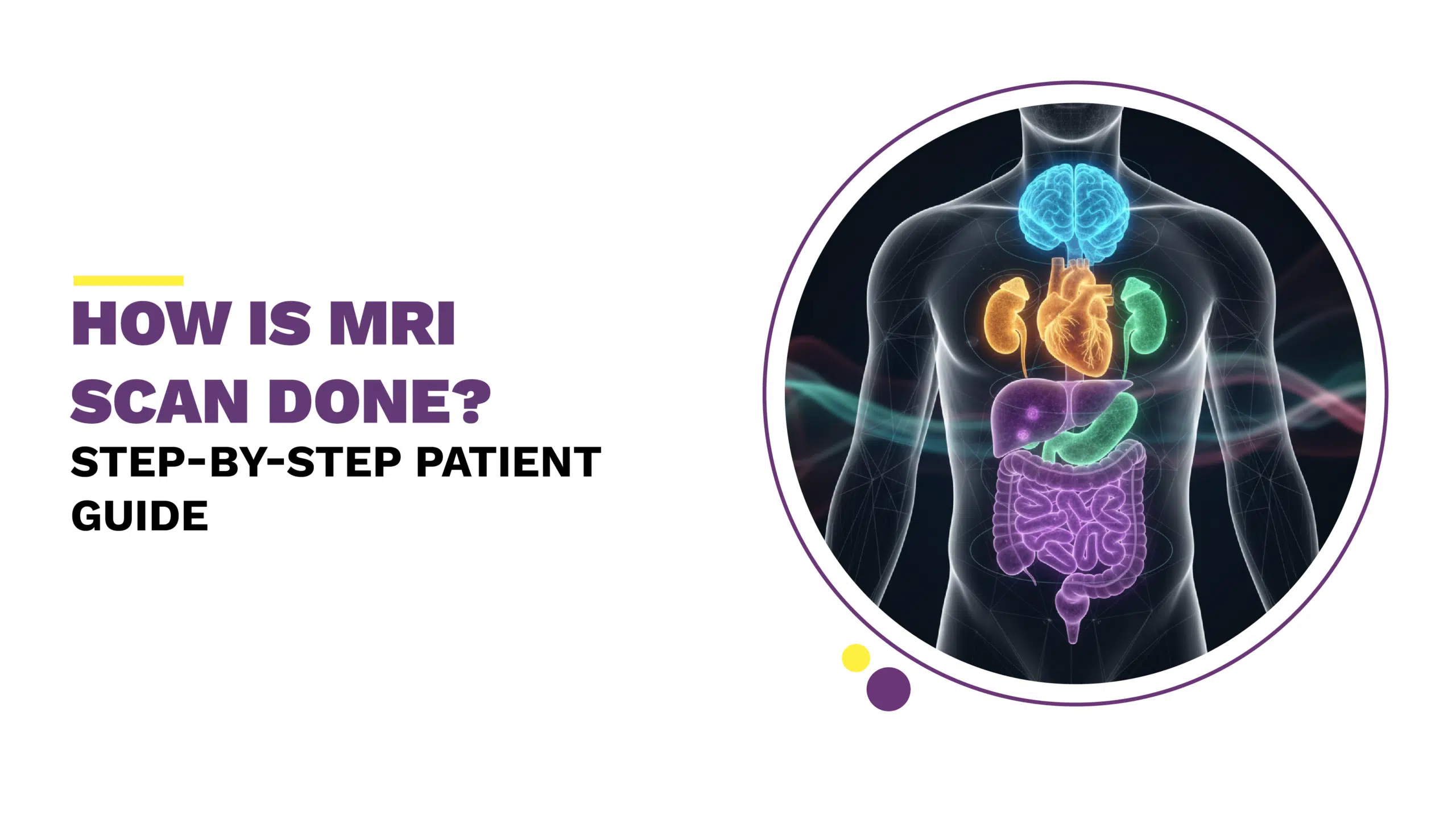
Understanding MRI: Why It Matters in Diagnosing Disease
MRI uses strong magnetic fields and radiofrequency signals to create detailed pictures of organs, soft tissues, nerves, blood vessels, and joints. Unlike X-ray or CT scans, MRI does not use ionizing radiation. This makes it especially valuable when evaluating neurological conditions, spine disorders, musculoskeletal injuries, cardiac function, and many internal organ conditions.
At Images Diagnostic Center, patients benefit from advanced MRI systems, including 3 Tesla MRI scanners, which provide higher clarity, faster imaging times, and enhanced precision for complex cases such as neurological evaluation, tumor characterization, and joint imaging.
How Is an MRI Scan Done? Inside the Room, Step by Step
MRI is performed in a controlled environment managed by specialized technologists and interpreted by board-certified radiologists. Here is what typically happens:
1. Patient Registration and Safety Screening
Before the scan, patients complete a safety form that screens for:
- Pacemakers.
- Metallic implants.
- Artificial joints.
- Surgical clips.
- Claustrophobia.
- Previous surgeries.
This step ensures safety because the MRI magnet interacts with metals and certain implanted devices. Experienced technologists at Images Diagnostic Center review these forms and ask follow-up questions if needed.
2. Preparation Before Entering the MRI Suite
Patients may be asked to:
- Change into medical gowns.
- Remove jewelry, watches, credit cards, or hairpins.
- Leave electronic devices outside
Some patients require contrast injection depending on the diagnostic purpose. Contrast helps highlight blood vessels, inflammation, or lesions when needed.
3. Positioning Inside the MRI Machine
Once inside the MRI room, the technologist helps the patient lie on the scanning table. Soft cushions support the head, knees, or spine as needed. For spinal or brain scans, head coils or body coils are placed around the region to enhance signal detection.
Patients are given a buzzer to call the technologist if needed and headphones to lessen noise.
4. The Imaging Process
Once the table moves into the magnet, the MRI system begins acquiring images. The patient hears tapping or vibrating sounds, normal and expected.
Remaining still is critical. Even slight movement may blur images, requiring repeat scans.
At Images Radiology Center, advanced noise-reduction MRIs and scan-time optimization improve comfort, helping patients tolerate the procedure more easily.
5. Contrast-Enhanced Imaging (if required)
If contrast is needed, a technologist will inject it halfway through the scan. Contrast agents provide additional diagnostic information for tumors, vascular conditions, and inflammatory disease.
6. Scan Completion and Review
When imaging concludes, the table moves out and coils are removed. Patients resume normal activities unless their physician recommends observation. Results are then analyzed by a radiologist who prepares an expert report for the referring physician.
What Makes 3 Tesla MRI More Accurate?
A 3 Tesla MRI scan in Kuwait provides higher magnetic strength than conventional 1.5T machines, allowing:
- Higher resolution images.
- Shorter scan time.
- Better visualization of brain and spinal structures.
- Enhanced musculoskeletal imaging.
- Improved vascular MRI without contrast.
These advantages support earlier diagnosis and more accurate treatment planning. At Images Diagnostic Center, 3T MRI is routinely used for neurological disorders, orthopedic injuries, liver imaging, abdominal pathology, and complex cardiac studies.
What Does an MRI Help Diagnose?
MRI is not limited to one specialty. It assists in diagnosing:
- Brain tumors, stroke, epilepsy.
- Multiple sclerosis or spinal disc disease.
- Ligament and tendon injuries.
- Liver fibrosis through elastography.
- Joint degeneration.
- Cardiomyopathy and heart function.
- Pelvic and abdominal pathology.
MRI is also important in oncology, where tumor size and progression must be monitored precisely.
The Patient Experience: Comfort, Noise, and Monitoring
Many first-time patients ask if MRI is painful. The scan itself is painless. Patients may feel:
- Slight warmth in the scanned area.
- Mild anxiety from enclosed spaces.
- Noise discomfort.
Technologists are present throughout the scan. Patients receive intercom communication, reminding them they are never alone inside the scanner. For anxious or elderly patients, Images Diagnostic Center offers open MRI scan options, which are more comfortable and less confining.
MRI vs CT Scan: When Is Each Preferred?
Both MRI and CT are important diagnostic tools, but they serve different purposes:
| MRI | CT Scan |
| Uses magnetic field | Uses X-rays |
| Best for soft tissue, brain, spine, joints | Best for bone, lung, trauma, vascular calcification |
| Longer scan time | Fast scan (seconds to minutes) |
| No radiation exposure | Low radiation dose |
How fast can CT scan results be obtained?
At Images, results are typically available within hours because reports are read by senior radiologists experienced in cross-sectional interpretation.
CT is particularly valuable for:
- Emergency stroke imaging.
- Trauma assessment.
- Lung opacity evaluation.
- Cardiac calcium scoring.
Whereas MRI is chosen for neurologic, muscular, spinal, and organ integrity imaging.
Ultrasound Imaging: The First-Line, Real-Time Diagnostic Tool
Ultrasound and Doppler scans are non-invasive, safe during pregnancy, and deliver real-time visualization.
They help evaluate:
- Abdominal organs.
- Blood vessels (Doppler).
- Thyroid, breast tissue.
- Pregnancy.
For soft tissue concerns, ultrasound frequently guides further testing, MRI or CT, when needed.
Breast Imaging and Mammography: Detecting Early Disease
A mammogram is one of the most efficient screening tools for breast cancer. Images Diagnostic Center provides digital mammography, improving detection for breast density and microcalcification analysis.
When combined with breast ultrasound or MRI for high-risk patients, early detection becomes far more likely.
Bone Density (DEXA Scan): Tracking Osteoporosis Risk
DEXA scan measures bone mineral density and is especially recommended for postmenopausal women, elderly patients, or individuals on long-term steroids.
Benefits include:
- Early fracture risk prediction.
- Monitoring osteoporosis treatment.
- Guiding calcium and vitamin D therapy.
Home Radiology: When Imaging Comes to You
Some patients, elderly, post-surgical, or mobility-limited, cannot visit imaging centers. Images GO, the home radiology service in Kuwait, offers portable X-ray and ultrasound imaging performed by certified technologists.
This mobile imaging for patients at home provides:
- Convenience.
- Professional reporting by radiologists.
- Reduced risk of hospital exposure.
This service is particularly valuable for orthopedic cases, bedridden patients, and chronic conditions.
Patient Preparation Tips for MRI
Patients often ask how they can prepare for their MRI appointment:
Before the scan:
- Eat normally unless instructed otherwise.
- Inform the team if pregnant or breastfeeding.
- Notify technologists of previous surgeries or implants.
During the scan:
- Remain still for best image quality.
- Communicate if anxious.
- Follow breathing instructions if given.
After the scan:
- Resume normal activity unless contrast after-care is required.
- Follow up with your physician to discuss results.
Imaging Done Right, When Accuracy Matters
Radiology is more than pictures, it is clinical decision support. Whether performing a 3 Tesla MRI, CT angiography, ultrasound, breast imaging, or home radiology, the expertise behind the machine determines diagnostic confidence.
With international accreditations, advanced technologies, and subspecialty radiology expertise, Images Diagnostic Center in Kuwait delivers precision imaging, compassionate care, and results that help physicians make informed treatment choices.
Accurate diagnostics are proactive healthcare, not reactive treatment. Choosing timely imaging allows early detection, monitoring of disease, and peace of mind.
FAQ Section
1. What is the difference between contrast and non-contrast MRI?
A non-contrast MRI assesses anatomy, injury, or inflammation using only magnetic signals. A contrast MRI uses gadolinium-based agents to highlight abnormal blood flow, tumors, infection, scarring, and post-surgical changes. Radiologists request contrast when evaluating suspected lesions or vascular conditions.
2. How does cardiac MRI evaluate heart function?
Cardiac MRI visualizes heart muscle, valves, chambers, and blood flow dynamics. It assesses:
- Pumping efficiency.
- Scar tissue.
- Congenital abnormalities.
- Myocarditis.
It is widely used for cardiomyopathy evaluation and complements echo and CT imaging.
3. Can MRI detect inflammation without contrast?
Yes. Modern 3 Tesla MRI scans can reveal edema, joint effusion, and nerve inflammation using specialized signal sequences. However, contrast may be required for full assessment of infection or tumors.
4. When is ultrasound better than MRI?
Ultrasound is preferred for:
- Pregnancy.
- Thyroid nodules.
- Breast evaluation.
- Doppler vascular studies.
- Abdominal organ screening.
It provides real-time imaging, while MRI is selected when deeper internal or neurological detail is needed.
5. How often should bone density be monitored?
Patients with osteoporosis, steroid use, postmenopausal status, or fracture history typically undergo DEXA scans every 1–2 years. Frequency is determined by clinical risk and treatment changes.
6. Do children require special MRI preparation?
Children may require:
- Sedation (for movement control).
- Child-friendly explanation.
- Noise protection.
- Parental presence during setup.
At specialized units like Images Diagnostic Center, pediatric scanning is supervised by trained technologists and radiologists.
Get Your Scan with Confidence – Book at Images Diagnostic Center
- MRI (3 Tesla & Open MRI)
- CT Scan
- Ultrasound & Doppler
- Mammography
- DEXA Bone Density
- Home Radiology (Images GO)
Contact Us
Jabriya | Hawally | Salmiya
(+965) 1899 888
info@imagesforhealth.com
Your health deserves accurate answers, and they begin with high-quality imaging interpreted by expert radiologists.