
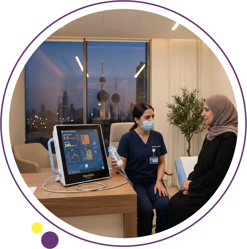
For decades, the only reliable way to know how much scarring, or fibrosis, had developed in a patient’s liver was a biopsy: a needle inserted through the skin to extract a small sample of tissue for laboratory analysis. FibroScan changed that equation. This non-invasive device measures liver stiffness directly through the skin in about ten minutes, with no needles, no sedation, and no waiting days for a pathology report.
In this article, we explain exactly how FibroScan works, what the kilopascal measurement actually represents, how the separate CAP score grades fat content in the liver, who should consider this test, and how it compares to liver biopsy and other imaging methods used to assess liver health.
What Is a FibroScan?
FibroScan is the trade name for a technique called transient elastography, a specialised ultrasound-based device that measures the stiffness of liver tissue. The device uses a probe placed against the skin over the liver, similar in appearance to a standard ultrasound probe, but instead of forming an image, it generates a low-frequency mechanical vibration that travels through the liver tissue and measures how fast that vibration propagates.
The core physical principle is straightforward: stiffer tissue transmits mechanical waves faster than soft tissue. A healthy, supple liver transmits the vibration slowly. A liver with significant scarring, or fibrosis, has become firmer and transmits the same vibration considerably faster. By measuring this speed precisely, FibroScan converts a physical property, tissue stiffness, into a clinically meaningful number without ever penetrating the skin. Images Diagnostic Center in Kuwait offers this test as part of a comprehensive approach to liver health assessment.
How Liver Stiffness Is Measured in Kilopascals
The result of a FibroScan examination is expressed in kilopascals (kPa), a unit of pressure that in this context reflects the elastic stiffness of liver tissue. A healthy liver typically measures in the range of roughly 2 to 7 kPa. As fibrosis develops and progresses, this number rises steadily: mild fibrosis might measure in the high single digits, moderate fibrosis in the low teens, and advanced fibrosis or cirrhosis often measures well above 12 to 14 kPa, with some cirrhotic livers measuring 20 kPa or higher.
This numerical scale allows clinicians to track fibrosis progression or regression over time with a precision that qualitative descriptions cannot match. A patient monitored annually can compare their kPa value year over year to see whether treatment is working, whether the disease is stable, or whether it is advancing and requires more aggressive intervention. Clinical validation studies have established these kPa thresholds against liver biopsy results across large patient populations, giving the test a strong evidence base. The FibroScan service at Images provides this precise numerical tracking for patients in Kuwait.
The CAP Score: A Separate Measurement of Fat Content
What makes modern FibroScan devices particularly useful is that they measure two entirely separate things in a single session. Alongside the stiffness measurement in kPa, the device also calculates a Controlled Attenuation Parameter, or CAP score, measured in decibels per metre (dB/m). While kPa reflects scarring, CAP reflects the amount of fat accumulated within liver tissue, addressing an entirely different biological question.
The CAP score works by measuring how much the ultrasound signal weakens, or attenuates, as it passes through liver tissue. Fat droplets within liver cells scatter and absorb ultrasound energy more than normal liver tissue does, so a fattier liver produces a higher CAP score. A CAP score below roughly 238 dB/m generally indicates no significant fat accumulation, while progressively higher values indicate mild, moderate, or severe steatosis. Having both kPa and CAP from the same non-invasive session gives a remarkably complete snapshot: how much fat is present, and how much scarring has resulted from it. This dual output is particularly valuable when assessing fatty liver disease, where fat accumulation and fibrosis often coexist but progress at different rates. For a comprehensive look at how fat accumulation and fibrosis relate to each other across the stages of liver disease, our detailed guide on fatty liver disease covers the full clinical picture.
What Happens During a FibroScan Examination
A FibroScan examination is remarkably simple from the patient’s perspective. You lie on your back with your right arm raised behind your head, similar to the positioning used for a standard abdominal ultrasound. The technologist locates an appropriate intercostal space, between the ribs on the right side, and places the probe against the skin at this location.
The device then takes a series of individual measurements, typically ten or more, at slightly different depths and positions within the same general area. Each measurement takes only a second or two, and the machine displays immediate feedback on measurement quality. The entire session, including positioning and the full set of measurements, is usually completed within ten minutes. There is no injection, no contrast material, no radiation, and no sedation required, meaning patients can return to normal activity immediately afterward. Fasting for a few hours beforehand is typically recommended, as a full stomach can affect measurement accuracy by altering liver position and blood flow.
Who Should Consider a FibroScan?
FibroScan is most valuable for patients with an established or suspected liver condition where the degree of fibrosis materially changes management decisions. This includes patients with fatty liver disease (both alcoholic and non-alcoholic forms), chronic viral hepatitis B or C, and other chronic liver conditions where ongoing inflammation could be driving progressive scarring.
It is particularly useful for patients with metabolic risk factors including obesity, type 2 diabetes, and elevated liver enzymes on blood tests, where an ultrasound may have already suggested fat accumulation but the clinical question of how much fibrosis has developed remains unanswered. Current clinical guidance increasingly recommends FibroScan as a first-line non-invasive assessment before considering liver biopsy, reserving biopsy for cases where the FibroScan result is ambiguous or where additional diagnostic information is specifically needed. Patients being monitored for treatment response, whether through weight loss, diabetes control, or antiviral therapy, also benefit from serial FibroScan measurements to track whether their liver is improving over time.
FibroScan Versus Liver Biopsy
Liver biopsy has long been considered the reference standard for assessing fibrosis because it allows direct microscopic examination of liver tissue, revealing not just the degree of scarring but also cellular detail about inflammation, fat droplet distribution, and other pathological features that imaging cannot capture. However, biopsy is invasive, carries a small but real risk of bleeding and pain, and only samples a tiny fragment of the liver, roughly one fifty-thousandth of the total organ, which introduces sampling variability since fibrosis is not always distributed evenly.
FibroScan, by contrast, assesses a substantially larger volume of liver tissue with each measurement and carries no procedural risk at all. Its correlation with biopsy-confirmed fibrosis stages is strong, particularly at the extremes of the spectrum: distinguishing no fibrosis from advanced fibrosis or cirrhosis is highly reliable. The main limitation is in the intermediate range, where the correlation between kPa values and precise fibrosis stage is less exact, and some patients, particularly those with significant obesity or ascites, may have technically difficult or unreliable measurements. For most patients today, FibroScan serves as the first and often only assessment needed, with biopsy reserved for cases requiring additional diagnostic clarity that imaging alone cannot resolve.
FibroScan Compared to Standard Ultrasound and MRI
Standard abdominal ultrasound is excellent at detecting the presence of fat within the liver, visible as increased brightness of liver tissue compared to the adjacent kidney, but it cannot quantify fibrosis with any precision and cannot reliably distinguish mild fibrosis from more advanced stages. FibroScan fills exactly this gap, providing the quantitative fibrosis assessment that standard ultrasound cannot offer.
MRI-based techniques, particularly Magnetic Resonance Elastography (MRE), offer an alternative method of measuring liver stiffness with excellent accuracy and the ability to assess the entire liver rather than a localised sample, but at substantially higher cost and with longer scan times than FibroScan. MRI also remains valuable when FibroScan results are inconclusive or when additional anatomical detail is needed, such as evaluating focal liver lesions alongside diffuse fibrosis. For most routine fibrosis monitoring, however, FibroScan offers the best balance of accuracy, speed, and accessibility. CT scanning plays a complementary role primarily in assessing complications of advanced liver disease, such as portal hypertension or ascites, rather than in fibrosis quantification itself.
Limitations and Considerations
While FibroScan is highly reliable for most patients, certain factors can affect measurement quality. Significant obesity can make it more difficult for the mechanical vibration to travel reliably through tissue, though modern devices include an XL probe specifically designed to improve accuracy in patients with a higher body mass index. Ascites, the accumulation of fluid in the abdominal cavity, can also interfere with accurate measurement since the vibration does not transmit reliably through fluid.
Recent food intake, active inflammation from acute hepatitis, and congestive states affecting the liver can also transiently elevate stiffness readings independent of true fibrosis, which is why fasting beforehand and interpreting results in clinical context both matter. Professional guidelines recommend interpreting FibroScan results alongside blood tests and clinical history rather than as an isolated number, ensuring the most accurate overall picture of liver health. The team at Images incorporates this comprehensive interpretation approach for every patient undergoing this examination.
Frequently Asked Questions
Is FibroScan painful?
No, FibroScan is completely painless. Patients feel a brief, mild vibration or tapping sensation against the skin during each measurement, similar to a gentle pluck, but there is no needle, no incision, and no discomfort comparable to a biopsy. Most patients describe the experience as similar to a routine ultrasound.
Do I need to fast before a FibroScan?
Yes, fasting for approximately two to three hours beforehand is typically recommended. Recent food intake increases blood flow to the liver and can temporarily elevate stiffness readings, potentially overestimating the true degree of fibrosis. Following the fasting instructions given by the imaging team helps ensure the most accurate result.
How often should FibroScan be repeated?
This depends on the underlying condition and its stability. Patients with mild, stable fatty liver disease might be reassessed annually, while those undergoing active treatment for a progressive condition, or those with more advanced fibrosis, may need more frequent monitoring, sometimes every six months, to track response to treatment or disease progression. Your physician will determine the appropriate interval based on your specific situation.
Can FibroScan replace a liver biopsy completely?
For the majority of patients being monitored for fatty liver disease or chronic viral hepatitis, FibroScan has largely replaced the need for routine biopsy. However, biopsy remains valuable in select cases, such as when FibroScan results are technically unreliable, when the diagnosis remains unclear despite imaging, or when detailed cellular information beyond fibrosis stage is specifically needed for treatment decisions.
Where can I get a FibroScan in Kuwait?
Images Diagnostic Center provides FibroScan examinations at its Jabriya and Salmiya branches in Kuwait, alongside complementary liver imaging including ultrasound, CT, and MRI. You can contact Images to arrange an appointment, whether following a referral or as part of proactive liver health screening.
A Simple Test With Substantial Clinical Value
FibroScan has transformed how liver fibrosis is assessed and monitored, replacing what once required an invasive biopsy with a ten-minute, needle-free examination that delivers precise, repeatable numerical results. Combined with the CAP score’s separate assessment of fat content, a single FibroScan session now answers two of the most clinically important questions in liver disease at once.
Images Diagnostic Center provides FibroScan and complementary liver imaging across two Kuwait branches:
To arrange a FibroScan examination, contact Images directly.