
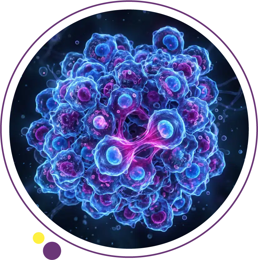
Every cancer, regardless of the organ it develops in or the treatment it ultimately requires, begins with a single cell that has crossed a line. That cell has accumulated enough genetic damage to break free from the rules that govern how normal cells behave: when to grow, when to stop growing, and when to die. Understanding what cancer cells are, how they differ from healthy cells, and how they spread throughout the body is the foundation for understanding why cancer behaves the way it does and why imaging plays such a central role in tracking its behaviour.
In this article, we will explain the biology of cancer cells in accessible but accurate terms, covering how DNA mutations transform normal cells into cancer cells, the hallmarks that define cancer cell behaviour, how tumors grow their own blood supply, how cancer spreads to distant organs, and critically, how modern imaging detects and monitors these processes at every stage.
How Normal Cells Become Cancer Cells
Every cell in the human body contains a complete copy of the genome, approximately three billion base pairs of DNA encoding the instructions for all cellular functions. Within that genome are proto-oncogenes that promote cell growth and division, tumour suppressor genes that act as brakes on cell division, and DNA repair genes that correct replication errors before they accumulate into permanent mutations. Normal cell growth is a tightly regulated process: cells receive a signal to divide, complete division, and then wait for the next signal.
Cancer begins when mutations occur in one or more of these regulatory genes. A single mutation is rarely sufficient to produce a cancer cell. Instead, cancer develops through a process of clonal evolution: a cell accumulates one mutation, its daughter cells inherit that mutation and acquire additional ones over time, and eventually a clone emerges with enough genetic alterations to sustain uncontrolled growth. Research on this process has shown that most common cancers require the accumulation of at least four to seven key driver mutations before frank malignancy develops, which is why cancer is predominantly a disease of older age when DNA has had decades to accumulate replication errors. This slow developmental timeline is precisely why early detection through imaging at Images Diagnostic Center in Kuwait can find cancer before it has progressed to an untreatable stage.
The Hallmarks of Cancer Cells
In landmark work first published in 2000 and updated in subsequent years, cancer biologists Robert Hanahan and Douglas Weinberg identified a set of functional capabilities that cancer cells must acquire to become malignant tumors. These hallmarks of cancer have become the conceptual framework through which cancer biology is understood and through which new treatments are developed. Understanding them helps explain why cancer is so difficult to eliminate once established.
The core hallmarks include: self-sufficiency in growth signals, meaning cancer cells produce their own growth-promoting signals rather than waiting for external signals; resistance to growth-inhibitory signals, meaning the normal brake mechanisms are disabled; evasion of programmed cell death (apoptosis), meaning the cell death pathway that would normally eliminate damaged cells is bypassed; limitless replicative potential, meaning cancer cells do not reach the normal limit on the number of times they can divide; the ability to sustain angiogenesis (growing a new blood supply); and the capacity to invade local tissue and spread to distant sites. Two additional enabling characteristics include genetic instability that accelerates mutation accumulation, and inflammation that provides growth signals and immune evasion. The CT and MRI services at Images can detect the structural consequences of these cellular processes, from the appearance of a mass to signs of invasion and spread.
How Cancer Cells Bypass the Cell Cycle
The cell cycle is the sequence of phases a cell passes through when it divides: growth phases, a DNA synthesis phase where the genome is duplicated, and a mitotic phase where the cell divides into two daughter cells. At several points within this cycle are checkpoints where the cell’s internal machinery verifies that everything is in order before proceeding. If DNA is damaged or incompletely replicated, the checkpoint halts the cycle and either allows time for repair or triggers apoptosis if the damage is irreparable.
Cancer cells disable these checkpoints through mutations in genes including TP53, RB1, and the CDKN family of cyclin-dependent kinase inhibitors. TP53, encoding the p53 protein, is the most frequently mutated gene in human cancer and is sometimes called the guardian of the genome because of its central role in responding to DNA damage and triggering apoptosis. When p53 function is lost, cells with damaged DNA continue to divide and accumulate further mutations rather than being eliminated. This drives the rapid genetic instability that characterises aggressive cancers. The imaging services at Images can assess the macroscopic consequences of these processes, detecting the masses that accumulate as these cell populations expand.
Angiogenesis: How Cancer Cells Grow Their Own Blood Supply
A tumor cannot grow beyond approximately two millimetres in diameter without establishing a blood supply to deliver oxygen and nutrients. Beyond this size, cells in the tumor interior become hypoxic (oxygen-deprived) and begin to die. To overcome this limitation, cancer cells exploit the normal process of blood vessel formation, known as angiogenesis, by secreting vascular endothelial growth factor (VEGF) and other pro-angiogenic signals. These signals recruit endothelial cells from nearby vessels to sprout new capillaries into the growing tumor mass.
The blood vessels that cancer cells recruit are structurally abnormal: they are leaky, irregularly branched, and poorly regulated. This abnormal vascularity has both biological consequences, including variable drug delivery within the tumor, and imaging consequences that are diagnostically useful. The enhancement pattern of a tumor after intravenous contrast administration on CT or MRI reflects this abnormal vascularity. Contrast-enhanced imaging identifies the territory of abnormal vessel growth, which corresponds to viable tumor, and areas of non-enhancement may indicate central necrosis. This is why contrast-enhanced CT and MRI at Images are so important for tumor characterisation rather than non-contrast studies alone.
How Cancer Spreads: The Metastatic Process
Metastasis, the spread of cancer from the primary site to distant organs, is responsible for approximately ninety percent of cancer deaths and represents the most clinically significant consequence of the biological changes in cancer cells. The metastatic process is complex and involves multiple steps, each of which represents a potential barrier to successful spread. The fact that metastasis occurs at all reflects how profoundly cancer cells have altered their biology compared to normal cells.
Cancer cells must first invade through the basement membrane and extracellular matrix surrounding the primary tumor. They achieve this by producing matrix metalloproteinases (MMPs), enzymes that digest structural proteins in the tissue architecture. Once free of the primary site, cancer cells enter blood vessels or lymphatics (intravasation), travel to distant sites, exit the circulation (extravasation), and then establish a micrometastatic colony in the new organ. The vast majority of circulating cancer cells are destroyed by immune mechanisms, physical trauma, or failure to find a permissive microenvironment. Only a small fraction successfully establishes metastatic deposits. Understanding where cancers commonly spread helps explain the staging imaging used after diagnosis, which at Images includes CT of the chest, abdomen, and pelvis, and brain MRI when intracranial metastases are possible.
Common Sites of Cancer Spread and Why They Occur
Different cancers have characteristic patterns of metastatic spread that are not random. These patterns reflect the anatomy of venous drainage, the biology of the cancer cells, and the molecular properties of the target organ that make it either permissive or non-permissive for metastatic colonisation. Colon cancer spreads to the liver via the portal venous system, which drains the bowel directly into the liver, making hepatic metastases the most common distant site in colorectal cancer. Lung cancer spreads to the brain, bone, liver, and adrenal glands. Breast cancer spreads to bone, liver, lung, and brain. Prostate cancer has a particular predilection for bone metastases.
These organ-specific tropisms were captured in the nineteenth-century concept of seed and soil, which holds that cancer cells (seeds) can only establish metastases in organs (soil) whose microenvironment provides the right conditions for growth. This concept remains valid today and continues to inform both biological research and clinical staging decisions. CT staging of colon cancer focuses on the liver and lungs as the highest-priority metastatic sites. Bone scan or whole-body MRI evaluates skeletal metastases in prostate and breast cancer. Brain MRI examines intracranial metastases in lung and breast cancer. These organ-targeted imaging protocols at Images are designed around the known metastatic biology of each cancer type. For a practical example of how this staging process works in a specific cancer type, our article on colon cancer explains how staging imaging is organised for colorectal disease.
Genetic Mutations in Cancer: Driver vs Passenger
Modern genomic sequencing of cancer tissue has revealed that cancer cells typically contain hundreds to thousands of mutations, far more than the handful of critical driver mutations needed to initiate and sustain malignancy. Researchers distinguish between driver mutations, which confer a growth advantage that contributes to cancer development, and passenger mutations, which are accumulated DNA damage events that have no functional consequence. Identifying which mutations are drivers and which are passengers is the central challenge of cancer genomics and the foundation of targeted therapy development.
The most therapeutically important driver mutations include KRAS, EGFR, ALK, BRAF, HER2, and TP53 across different cancer types. When a cancer carries an actionable driver mutation, a targeted drug that inhibits the protein encoded by that mutated gene can produce dramatic and prolonged responses in some patients. The molecular profiling of biopsy tissue has become mandatory in several cancer types to determine which targeted agents may be effective. Imaging supports this by identifying the optimal site for biopsy and subsequently monitoring response to targeted therapy through serial CT or MRI studies.
The Tumour Microenvironment
A tumor is not simply a collection of cancer cells in isolation. It is an organised and complex tissue mass that includes cancer cells, stromal fibroblasts, endothelial cells forming blood vessels, and a diverse range of immune cells. The interactions between these components, collectively called the tumour microenvironment, profoundly influence how the cancer grows, how it responds to treatment, and whether it evades immune surveillance. Cancer cells actively manipulate their microenvironment to suppress immune attack, recruit stromal support, and promote angiogenesis.
The tumour microenvironment is the target of immunotherapy: checkpoint inhibitor drugs work by releasing immune cells from the suppressive signals that cancer cells produce, allowing the immune system to recognise and kill cancer cells that were previously invisible to it. The clinical success of immunotherapy in cancers with high tumour mutation burden and specific immune infiltration patterns reflects the importance of the microenvironment in cancer biology. These treatments are monitored by serial imaging, and the response patterns can look different from chemotherapy responses on CT, sometimes producing initial apparent growth before regression. Understanding this on CT imaging at Images requires familiarity with these immunotherapy-specific response criteria. The Images health blog continues to cover these evolving topics for patients and clinical teams in Kuwait.
How Imaging Tracks Cancer Cell Growth and Spread
Imaging cannot see individual cancer cells, but it can detect the macroscopic consequences of cancer cell activity with high sensitivity and spatial resolution. CT scanning detects mass lesions based on their density and enhancement characteristics, measures their size with precision, identifies lymphadenopathy reflecting regional spread, and detects distant metastases across multiple organ systems in a single examination. MRI adds superior soft tissue contrast, enabling detailed characterisation of tumor margins, invasion depth, bone marrow involvement, and intracranial disease. PET-CT adds functional metabolic information by identifying areas of elevated glucose uptake characteristic of cancer cells, and can detect disease not visible on anatomical CT alone.
Serial imaging is the standard method for monitoring treatment response, comparing the size and number of measurable lesions at baseline with follow-up measurements at defined time points after treatment. The RECIST criteria (Response Evaluation Criteria in Solid Tumors) provide a standardised framework for classifying tumors as responding, stable, or progressing based on CT measurements. This systematic approach to response assessment is the backbone of cancer treatment monitoring in clinical oncology worldwide and depends entirely on the quality and consistency of the imaging studies. The CT and MRI services at Images Diagnostic Center in Kuwait provide the imaging quality and consistency needed to support this monitoring pathway reliably.
Frequently Asked Questions
Are cancer cells alive?
Yes, cancer cells are living cells. They grow, divide, consume nutrients, produce energy, and communicate with their environment. The critical difference from normal cells is that they have escaped the regulatory constraints that govern normal cell behaviour. Cancer cells are not foreign organisms invading the body; they are the body’s own cells that have undergone genetic transformation and now behave in ways that are harmful to the organism as a whole.
How quickly do cancer cells grow?
Growth rates vary enormously between cancer types and between individual tumors. Some cancers, like certain thyroid cancers and some prostate cancers, grow so slowly that they may never become clinically significant within a patient’s lifetime. Others, like small cell lung cancer and some leukaemias, can double in size within weeks and require immediate treatment. The doubling time of a tumor also changes as it grows, with early cancer cells often dividing faster than later ones when hypoxia and nutrient limitations slow growth. Imaging response to treatment reflects these growth kinetics.
Can the immune system destroy cancer cells?
Yes, and it does so constantly. Immune surveillance is the process by which the immune system recognises and eliminates abnormal cells before they develop into clinical cancer. Most cancer-initiating cells are destroyed before they can establish a growing tumor. The cancers that ultimately develop are those that have successfully evaded immune surveillance through multiple mechanisms including downregulation of cell surface molecules that mark them for immune attack, and production of immunosuppressive factors that disable nearby immune cells. Immunotherapy works by removing these evasion mechanisms and re-enabling effective immune attack.
How does imaging help detect cancer cells?
Imaging detects the structural and functional consequences of cancer cell accumulation rather than individual cells. A tumor large enough to be visible on CT or MRI contains millions of cells. The imaging characteristics that distinguish cancer from normal tissue include abnormal density or signal intensity, irregular margins, contrast enhancement from abnormal vascularity, and associated changes in surrounding tissue. PET-CT detects areas of elevated glucose metabolism, which is a metabolic hallmark of cancer cells that is visible even before a structural mass is apparent on CT. At Images, contrast-enhanced CT provides the quality needed for these diagnostic distinctions.
Is every tumour a cancer?
No. A tumour simply means an abnormal growth or mass of tissue. Tumours are classified as benign, which are non-invasive and do not metastasise, or malignant (cancer), which invade surrounding tissue and can spread to distant sites. Benign tumours can still cause symptoms by pressing on adjacent structures and may need treatment, but they do not carry the systemic risk of malignant tumours. The distinction between benign and malignant requires histopathological analysis of a tissue sample, and imaging can suggest but not definitively establish this distinction.
Understanding Cancer Biology Improves Every Patient’s Journey
The biology of cancer cells explains why certain cancers respond to specific treatments, why imaging is so central to monitoring cancer behaviour, why early detection before metastasis is so critical to outcomes, and why the molecular profile of a tumor matters as much as its anatomical location. Patients who understand these principles are better equipped to engage with their clinical teams, to understand what each imaging study is looking for, and to appreciate why the management of cancer has become such a complex and individualised field.
Images Diagnostic Center provides CT scanning and MRI across three Kuwait branches to support cancer detection, staging, and treatment monitoring:
To arrange imaging as part of cancer evaluation or monitoring in Kuwait, contact Images directly.