

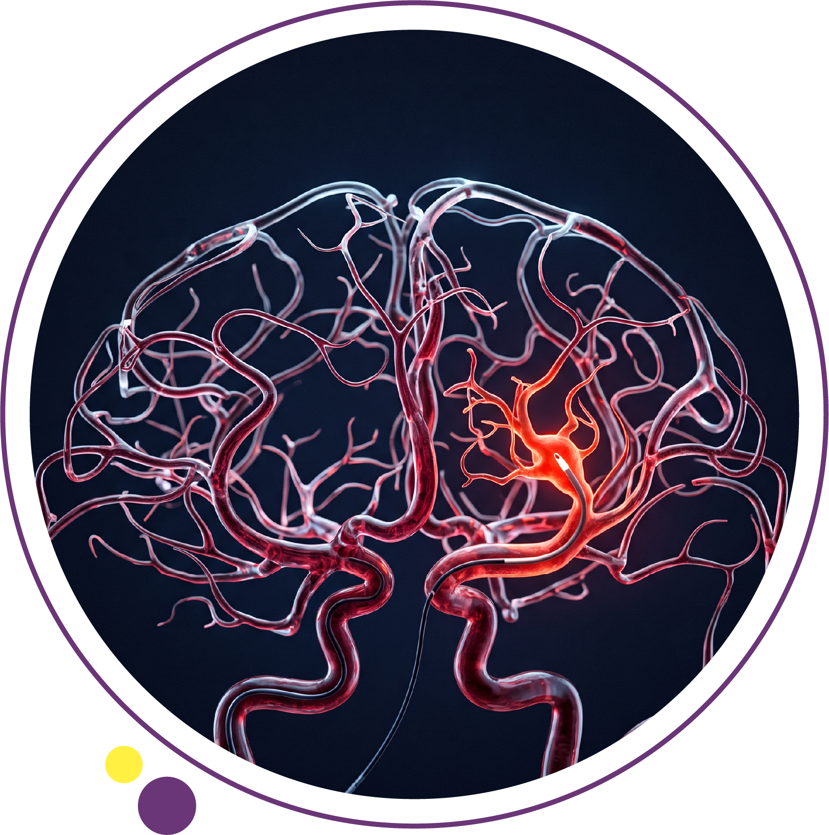
Interventional radiology (IR) uses medical imaging to guide treatments through small skin openings, often avoiding the need for a large surgical incision. A specialist may guide a thin catheter through a blood vessel, place a needle into a precise target or use energy to destroy abnormal tissue. The imaging equipment provides a road map while the procedure is being performed.
IR includes treatments for blocked arteries, internal bleeding, selected tumors and other conditions. It is different from a standard diagnostic CT, MRI or ultrasound examination, although those scans often help doctors plan an intervention. In Kuwait, IMAGES Diagnostic Center provides CT, MRI and ultrasound imaging that may support clinical evaluation. Its two operating branches are:
The procedures discussed below are general examples of interventional radiology, not a claim that IMAGES performs each treatment. Procedure availability should be confirmed with the treating specialist or hospital.
What is interventional radiology and how does it differ from surgery?
Interventional radiologists are physicians trained to diagnose disease and perform image-guided procedures. They use real-time X-rays, fluoroscopy, ultrasound or CT to reach a target while minimizing damage to surrounding tissues. Compared with some open operations, selected procedures can involve smaller incisions and a shorter recovery period; outcomes vary by condition and individual risk.
Diagnostic radiology focuses on producing and interpreting images. Interventional radiology uses imaging to guide needles, wires, tubes or devices to perform an action, such as opening a vessel or draining infected fluid. Conventional surgery remains essential for many conditions, and IR is not automatically safer or more effective in every case.
The Society of Interventional Radiology describes IR as a specialty that pairs imaging with targeted minimally invasive care across many organs.
How does imaging guide an interventional radiology procedure?
Different imaging tools offer different advantages during treatment.
Ultrasound-guided interventional procedures
Ultrasound shows soft tissues and moving needles in real time without ionizing radiation. Clinicians can use it to guide some biopsies, fluid drainage procedures and blood vessel access. The best approach depends on whether the target is clearly visible on ultrasound and can be reached safely.
CT-guided needle procedures
CT guidance provides precise cross-sectional views of structures that are difficult to see on ultrasound, including some deep lung, abdominal and bone targets. A specialist may use CT to plan a biopsy, access a collection or guide selected tumor treatments. CT uses X-rays, so the procedure team manages radiation exposure.
Fluoroscopy-guided catheter procedures
Fluoroscopy produces live X-ray images that allow a physician to advance thin wires and catheters through vessels. Some procedures require contrast material to show the vascular pathway. This imaging is commonly used for embolization, angioplasty and stent placement.
MRI also has specialized guidance roles, but it is not the standard imaging tool for every intervention. The choice reflects the target, clinical urgency, equipment and specialist expertise.
What procedures does interventional radiology perform?
IR is a broad field. The most familiar procedures fall into several groups, each intended to solve a different clinical problem.
Angioplasty and stenting for narrowed blood vessels
In angioplasty, a physician advances a balloon catheter into a narrowed blood vessel and inflates it to improve the opening. A stent, a small mesh tube, may be inserted to help keep the vessel open. These procedures are used in selected peripheral vascular conditions and other anatomical sites; candidacy depends on symptoms, imaging and the pattern of disease.
Embolization to block a targeted blood supply
Embolization deliberately blocks a chosen blood vessel using tiny particles, coils or other agents delivered through a catheter. It can help control certain types of internal bleeding, treat selected aneurysms or reduce the blood supply to a tumor or uterine fibroid.
For example, uterine fibroid embolization (UFE) aims to reduce blood flow to symptomatic fibroids so they shrink over time. It is one of several treatment options and requires consultation about fertility goals, symptoms and alternatives. The RadiologyInfo guide to catheter embolization outlines common indications and safety considerations.
Image-guided biopsies to sample abnormal tissue
A biopsy uses a needle to collect cells or tissue from a suspicious area. CT or ultrasound guidance can help select a safe route to a lump or lesion. A pathologist examines the sample to provide a tissue diagnosis. Imaging may locate a suspicious mass, but it cannot replace biopsy when tissue confirmation is needed.
Image-guided drainage of infected or trapped fluid
When an abscess or another fluid collection needs drainage, an interventional radiologist may insert a thin catheter through the skin into the target under imaging guidance. The catheter allows fluid to drain while treatment continues. Some collections still require surgery, and suspected infection may also require antibiotics and urgent medical care.
Tumor ablation using heat or cold
Ablation aims to destroy selected abnormal tissue with energy delivered through a probe. Options include radiofrequency or microwave heating and cryoablation, which uses extreme cold. These treatments can be considered for particular tumors according to location, size, organ function and the broader cancer plan. Ablation is not a universal replacement for surgery, radiotherapy or systemic cancer treatment.
Catheter-based treatments for blood clots
Selected cases of severe clot-related disease may be treated with catheter-directed clot removal or medication delivered near the blockage. Not every deep vein thrombosis or pulmonary embolism calls for an interventional procedure. Decisions depend on urgency, bleeding risk, symptoms and specialist guidelines.
Which conditions may benefit from interventional radiology?
Examples include selected cases of peripheral arterial disease, significant internal bleeding, uterine fibroids, certain tumors, deep abscesses, abnormal blood vessels and complications of venous disease. In some hospitals, IR teams also help with dialysis access and complex biliary or urinary drainage.
A condition may have multiple treatment pathways. For example, fibroids may be managed with monitoring, medication, embolization or surgery depending on symptoms and pregnancy plans. Cancer care may involve surgery, medication, radiation and targeted IR techniques, sometimes in combination.
A treatment is not chosen simply because it is less invasive. Doctors consider how likely it is to help, whether it can treat the entire problem and what follow-up is required.
How do you prepare for an interventional radiology procedure?
Preparation is more detailed than for many routine diagnostic scans because it depends on the treatment and type of anesthesia or sedation planned. The team will review your medical history, current medicines, allergies, past reactions to contrast, bleeding problems and kidney function when relevant.
You may need blood tests, updated imaging or instructions about eating and drinking. Tell the treating team if you take anticoagulants or antiplatelet medicines; never stop these drugs without explicit instructions. People who could be pregnant should tell the team before any X-ray-guided procedure. Ask whether someone needs to accompany you home and whether an overnight stay is expected.
What happens during an interventional radiology procedure?
The steps depend on the intervention, but the general process often includes:
- Clinical review: The team explains the goal, alternatives, risks and expected recovery, then obtains informed consent.
- Monitoring and pain control: Staff check vital signs, prepare the skin and provide local anesthetic, sedation or general anesthesia when needed.
- Image-guided access: The specialist makes a small opening or needle puncture and uses imaging to reach a vessel or organ.
- Treatment: A balloon, stent, embolic material, drainage catheter, biopsy needle or ablation probe is used according to the plan.
- Recovery and follow-up: The team checks for bleeding or complications and provides instructions on wound care, activity and further imaging.
Some procedures are done as day cases; others require a hospital admission. It is misleading to promise a fixed recovery time for all interventional radiology treatments.
What are the risks and limitations of interventional radiology?
Potential risks include bleeding, infection, injury to a blood vessel or nearby organ, pain and reactions to contrast or medication. Some procedures use X-ray guidance and involve radiation exposure. Certain patients face higher risks because of their underlying disease, blood-thinning medication or kidney problems.
IR may also be incomplete or unsuccessful, requiring another session, a different treatment or conventional surgery. Patients should ask about the expected benefit, alternatives, the likelihood of repeat treatment and how outcomes will be monitored. The relevant risk profile depends on the actual intervention, not just the label “minimally invasive.”
Diagnostic scans used before and after interventional radiology
Before an intervention, ultrasound and Doppler can assess soft tissues and blood flow, while CT can show detailed anatomy and the relationship of a target to nearby structures. MRI may help characterize tumors, uterine fibroids or other tissue abnormalities and assist treatment planning.
After a procedure, imaging can assess healing, residual disease, vessel patency or whether a collection has drained. Follow-up intervals vary by diagnosis and procedure. IMAGES offers ultrasound and Doppler as well as other diagnostic imaging; the referring physician chooses the required study and determines where any interventional treatment should take place.
Interventional radiology vs CT angiography: what is the difference?
CT angiography is usually a diagnostic test: contrast is injected into an arm vein and CT images map the vessels. Catheter angiography is more invasive: the physician places a catheter inside an artery and may be able to perform treatment during the same session. Interventional radiology refers to a much wider set of procedures, not just angiography.
For someone being assessed for vascular disease, a diagnostic test may be the first step. A separate treatment is considered only if the scan, symptoms and specialist assessment show that intervention is worthwhile.
Frequently asked questions about interventional radiology
Is interventional radiology the same as diagnostic radiology?
No. Diagnostic radiology mainly produces and interprets scans. Interventional radiology uses imaging to guide minimally invasive procedures that sample, drain or treat a specific condition.
Is interventional radiology a type of surgery?
Many IR procedures are minimally invasive medical treatments and are sometimes described as alternatives to open surgery. They still carry procedural risks and require trained specialists and appropriate facilities.
Are all interventional radiology procedures done without anesthesia?
No. Many use local anesthetic and sometimes sedation, while more complex procedures may require general anesthesia. The plan depends on the treatment and patient’s health.
How long does recovery take after interventional radiology?
Some patients return home the same day; others need monitoring or admission. Recovery can range from a short period to several weeks depending on the procedure.
Does interventional radiology use radiation?
Some procedures use fluoroscopy or CT and therefore involve ionizing radiation. Ultrasound-guided procedures do not. The team chooses imaging appropriate to the task and manages exposure.
Can interventional radiology treat cancer?
Certain tumor ablations and embolization techniques are part of interventional oncology, but not all cancers are suitable. Decisions are made within a broader cancer treatment plan.
Can a regular MRI or CT scan perform interventional treatment?
A standard diagnostic scan creates images but does not itself treat disease. Intervention requires trained clinicians, specific devices and a properly equipped procedural setting.
Diagnostic imaging for interventional care in Kuwait
Interventional radiology is an important treatment specialty, but diagnostic imaging and interventional treatment are not the same service. Ultrasound, CT and MRI often provide the information specialists need before or after a procedure.
If your clinician has requested diagnostic imaging as part of a treatment plan, contact IMAGES Diagnostic Center to confirm the correct examination and appointment at Jabriya or Salmiya. For an actual embolization, ablation, catheter treatment or other intervention, confirm availability directly with the treating specialist or hospital.
