

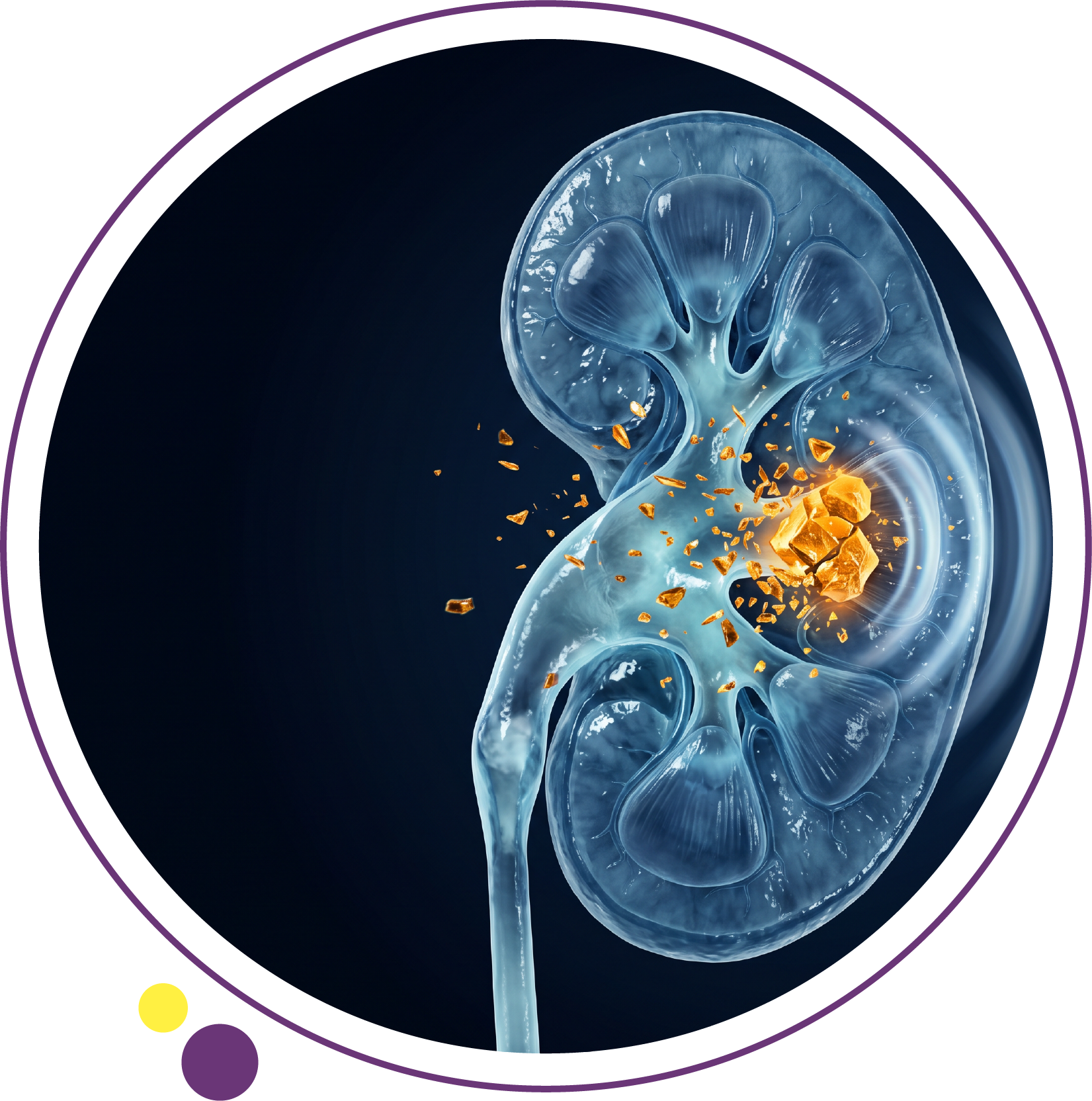
Kidney stones treatment depends on more than the size of a stone. Its location, the degree of blockage, symptoms, infection risk, kidney function, and whether the stone is growing all influence the safest choice. Some small stones pass through the urinary tract naturally; others need medication, shock wave treatment, endoscopy, or an operation to remove them.
Imaging is essential because pain does not reliably indicate stone size. A doctor may request ultrasound or a CT scan without contrast to locate the stone and assess obstruction. For physician-requested imaging in Kuwait, IMAGES Diagnostic Center has two active branches:
What Determines the Best Kidney Stones Treatment?
The two most important measurements are stone size and location. A 4 mm stone close to the bladder may pass more easily than a stone of the same size higher in the ureter. A non-obstructing stone inside the kidney may cause no symptoms, while a small stone blocking the ureter can cause severe pain.
A urologist also considers fever, urine test results, pain control, previous stones, pregnancy, kidney function, and whether the person has one functioning kidney. Treatment should follow a medical assessment; no chart based on millimeters alone can safely replace it.
Kidney Stones Treatment by Stone Size
Stones Under 5 mm: Observation and Pain Control
Many small ureteral stones pass without a procedure. If pain is controlled, there is no infection, and kidney function is not threatened, the clinician may recommend observation, appropriate pain medication, and follow-up imaging. The European Association of Urology (EAU) guidelines on urolithiasis report spontaneous passage in approximately three-quarters of ureteral stones smaller than 5 mm, with variation by location.
Passing a stone can take days to weeks. The absence of pain does not always prove that it has left the ureter, and scheduled follow-up helps confirm that obstruction has resolved.
Stones 5–10 mm: Medication or a Procedure May Be Needed
Stones in this range are less likely to pass as size increases, particularly higher in the ureter. Selected patients with distal ureteral stones larger than 5 mm may be offered an alpha-blocker, such as tamsulosin, to help passage; this use may be off-label depending on local rules. Such medication is not suitable for everyone and should only be used under medical advice.
If the stone is unlikely to pass, pain persists, or obstruction continues, ureteroscopy or shock wave lithotripsy may be considered. Location and stone density matter as much as the number on the CT report.
Stones 10–20 mm: More Likely to Need Active Treatment
Larger stones commonly require a procedure, but there is no single technique for every 10–20 mm stone. A specialist weighs whether it is in the kidney or ureter, its shape and density, anatomy, and expected chance of clearing all fragments. Shock wave treatment may be useful for some kidney stones, while ureteroscopy or flexible intrarenal surgery may provide a better option for others.
Stones Over 20 mm: Percutaneous Removal Is Often Preferred
For kidney stones larger than 2 cm, percutaneous nephrolithotomy (PCNL) is commonly recommended as the first-line option because it can remove a large stone burden more effectively than repeated shock wave sessions. Complex branching, or staghorn, stones often need this approach. The exact operation and recovery plan are individualized.
Kidney Stone Treatment Options Explained
Pain Relief and Fluids During Stone Passage
Nonsteroidal anti-inflammatory drugs are commonly used for renal colic when safe for the patient, but kidney disease, certain stomach problems, pregnancy, and other medical factors may limit their use. Other pain-control options can be considered by the treating clinician.
Drink enough fluid to avoid dehydration, but do not force excessive water during a severe obstructing episode. Drinking large amounts rapidly will not reliably push out a stuck stone and may worsen discomfort. A clinician should guide fluid intake when vomiting or kidney function is affected.
Medical Expulsive Therapy for Selected Ureteral Stones
Medical expulsive therapy uses medication to relax the ureter and improve the chances that certain stones pass. It is mainly considered for appropriately selected distal ureteral stones, especially those above 5 mm. It is not a substitute for urgent treatment when infection, uncontrolled pain, or deteriorating kidney function is present.
Shock Wave Lithotripsy (SWL)
Shock wave lithotripsy directs focused energy from outside the body at a stone to break it into smaller fragments. It generally does not require a surgical incision, but fragments must then pass through the urinary tract. Multiple sessions or another intervention may be needed. Success depends on size, position, density, and anatomy.
Ureteroscopy and Laser Lithotripsy
During ureteroscopy, a specialist passes a small scope through the urethra and bladder into the ureter, or sometimes farther into the kidney. The stone can be removed or broken using a laser. A temporary ureteral stent may be placed when appropriate. Recovery and discomfort vary according to the procedure and whether a stent is needed.
Percutaneous Nephrolithotomy (PCNL)
PCNL reaches the kidney through a small tract created through the back. It is typically used for larger or more complex stones. This is more invasive than shock wave treatment but can be more effective at clearing a large stone burden. Bleeding, infection, and other procedural risks require specialist discussion.
Can Medicine Dissolve Kidney Stones?
Most calcium-based stones cannot be dissolved with ordinary medication or lemon water. Some uric acid stones can be dissolved by carefully raising urine pH with prescribed medication and monitoring. Treatment depends on the stone’s composition, urine tests, and kidney function. Do not attempt to change urine acidity using unsupervised supplements.
What Imaging Is Needed Before Kidney Stone Treatment?
CT Scan Without Contrast for Stone Size and Location
Low-dose non-contrast CT is highly useful in suitable adults with suspected ureteral stones. It can define stone size, position, and density while showing signs of urinary blockage. These details influence the choice of observation, shock wave treatment, or endoscopic surgery. A referring doctor can request an appropriate CT scan at IMAGES.
Ultrasound for Selected Patients and Follow-Up
Ultrasound can show kidney stones and swelling caused by urinary obstruction. It avoids ionizing radiation and is commonly the preferred first imaging test during pregnancy and in children. Its ability to detect some small ureteral stones is lower than that of CT. IMAGES provides ultrasound and Doppler services for physician-referred examinations.
Why Stone Analysis and Urine Tests Matter
If a stone is passed or retrieved, analysis can identify its composition. Blood and urine tests assess kidney function, infection, and some metabolic factors. Patients with recurrent stones may benefit from a 24-hour urine collection to help plan prevention, rather than repeating the same treatment without investigating why stones form.
When Do Kidney Stones Require Emergency Treatment?
A stone that blocks urine drainage while an infection is present can become a life-threatening emergency. Urgent drainage using a ureteral stent or a nephrostomy tube may be needed, along with antibiotics. Definitive stone removal is generally delayed until the infection is controlled.
Go to emergency care for fever or chills with flank pain, inability to urinate, pain that remains severe despite prescribed treatment, persistent vomiting that prevents fluids, or signs of severe illness. Patients with a solitary kidney, pregnancy, or known kidney impairment should seek particularly prompt assessment.
How Long Does Recovery Take After Kidney Stone Treatment?
Recovery depends on the technique, the person’s health, and whether a stent is placed. Many people return to routine activity relatively quickly after an uncomplicated shock wave or ureteroscopic procedure, while recovery after PCNL may take longer. Mild bleeding or urinary discomfort can occur after procedures, but high fever, worsening pain, or inability to urinate requires urgent medical advice.
Follow-up is important because persistent fragments may remain after treatment. The clinician decides whether ultrasound, CT, or another examination is necessary to confirm clearance and assess kidney drainage.
How Can You Prevent New Kidney Stones?
Prevention should be tailored to stone type and the patient’s medical history. Common measures include adequate fluid intake, reducing excess salt, consuming a normal amount of dietary calcium rather than unnecessarily restricting it, and avoiding excessive animal protein. Hot weather, exercise, and outdoor work in Kuwait can increase fluid losses through sweating.
Certain patients may need medication to lower urine calcium, raise urine citrate, or manage uric acid. Medication is based on medical evaluation, not on an assumption that all stones have the same cause.
Frequently Asked Questions About Kidney Stones Treatment
Can a 4 mm kidney stone pass naturally?
Yes, many 4 mm ureteral stones pass without an intervention, particularly when they are near the bladder. Pain, obstruction, and infection still need assessment.
Does every 7 mm kidney stone need surgery?
No. Location and other factors matter. Some stones may pass with observation or selected medication, while others are better treated with a procedure.
Is laser treatment better than shock wave lithotripsy?
Neither is universally better. Ureteroscopy often clears certain stones more reliably in one procedure, while shock wave treatment can be less invasive for a suitable stone.
Do kidney stones always cause pain?
No. A non-obstructing kidney stone can remain silent. For more detail on flank pain, urine changes, and warning signs, see the site’s kidney stones symptoms guide.
Can lemon juice or drinking lots of water dissolve a stone?
Adequate hydration helps prevent many new stones, but most existing stones do not dissolve with water or lemon juice. Some uric acid stones can respond to prescribed urine alkalinization.
How does a doctor confirm that a stone has passed?
Passing and collecting the stone offers direct evidence, while follow-up imaging can assess whether a stone remains or blockage has resolved. Pain relief alone is not always enough.
Kidney Stone Imaging Before Treatment in Kuwait
Selecting kidney stones treatment begins with an accurate diagnosis, not an estimate based on pain. IMAGES Diagnostic Center provides the CT and ultrasound imaging that can help a referring urologist evaluate stone size, location, and urinary obstruction. The center does not replace the urologist who determines or performs treatment.
For a prescribed imaging examination at either active branch, contact IMAGES Diagnostic Center to confirm preparation and appointment availability.
