
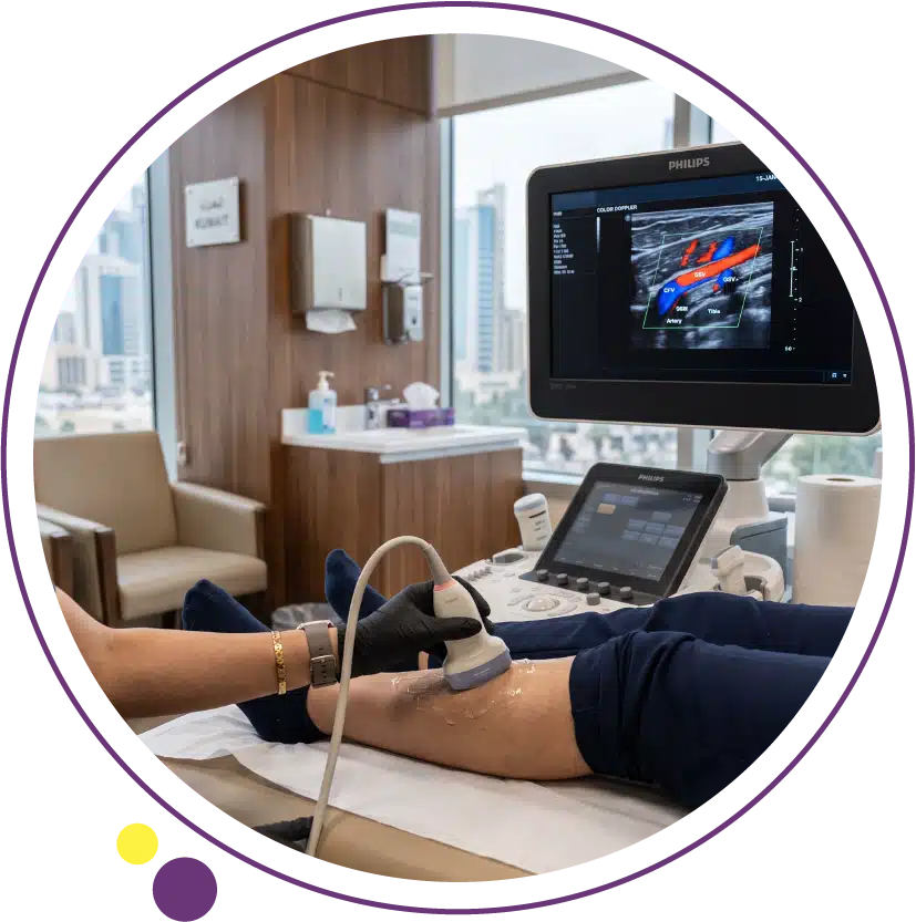
Standard ultrasound shows you what an organ or vessel looks like. Doppler ultrasound answers a different question entirely: how is blood actually moving through it right now. By measuring the speed and direction of blood flow using sound waves, without any radiation or injection involved, Doppler adds a functional dimension to imaging that a static picture alone cannot provide.
In this article, we explain the physical principle behind Doppler ultrasound, how colour mapping displays flow direction and speed on screen, the different types of Doppler used in clinical practice, and the common situations where this technique makes a genuine diagnostic difference.
The Physical Principle Behind Doppler Ultrasound
Doppler ultrasound relies on the same physical effect that causes a passing ambulance siren to sound higher in pitch as it approaches and lower as it moves away. Sound waves reflected off a moving object, in this case red blood cells travelling through a vessel, return to the probe at a slightly different frequency than they were sent, and the size of this frequency shift depends directly on how fast the blood is moving and in which direction relative to the probe.
The ultrasound machine captures this frequency shift and converts it into a precise measurement of blood flow velocity, all without any radiation, injection, or discomfort to the patient. This technology has become fundamental to vascular and obstetric imaging precisely because it adds functional flow information to the anatomical detail that standard ultrasound already provides. Doppler ultrasound at Images Diagnostic Center in Kuwait applies this principle across a wide range of clinical applications.
How Colour Maps Flow on Screen
Colour Doppler overlays a colour-coded map directly onto the standard greyscale ultrasound image, making blood flow immediately visible rather than requiring the interpreter to imagine it. By convention, flow moving toward the probe is typically displayed in red or orange tones, while flow moving away from the probe appears in blue tones, with brighter shades generally representing faster flow velocities.
This visual mapping allows a radiologist to instantly identify the direction blood is travelling through a vessel, spot areas of turbulent or disrupted flow that might indicate a narrowing or blockage, and confirm whether a structure of interest, such as a mass or an organ, has the blood supply that would be expected of it. Spectral Doppler, a complementary technique often used alongside the colour map, displays flow velocity as a waveform over time, allowing precise numerical measurement of peak velocities and flow patterns at a specific point within a vessel.
Types of Doppler Used in Clinical Practice
Colour Doppler provides the visual overview of flow direction and relative speed across an area, making it the first tool used to survey a vessel or region. Power Doppler is more sensitive to detecting the presence of flow, particularly in smaller vessels, though it does not show direction, making it useful for confirming blood supply to a small structure such as a testicle or an ovary. Spectral (pulsed-wave) Doppler provides the precise numerical velocity measurements needed for calculations such as resistance indices in pregnancy or velocity ratios used to grade the severity of an arterial narrowing.
Combining these Doppler modes within a single examination gives the complete picture: where flow is present, in which direction it travels, and precisely how fast it is moving, all gathered without radiation. The ultrasound service at Images uses this combined approach across its clinical applications in Kuwait.
Common Clinical Applications
In pregnancy, Doppler assesses blood flow through the umbilical cord and uterine arteries, helping identify pregnancies where the placenta may not be delivering blood flow as efficiently as expected. In vascular medicine, Doppler evaluates the leg veins for blood clots (deep vein thrombosis), assesses narrowing in the carotid arteries supplying the brain, and checks blood flow through arteries and grafts in the limbs.
Doppler also plays an important role in assessing organ transplants, confirming healthy blood supply to a transplanted kidney or liver, and in evaluating masses found on standard ultrasound, since the pattern of blood flow within a lesion can provide useful diagnostic clues about its nature. This breadth of application is exactly why Doppler has become a standard component added to many routine ultrasound examinations rather than a separate, rarely used test.
Frequently Asked Questions
Is Doppler ultrasound painful?
No, Doppler ultrasound is completely painless and uses the same probe and gel as a standard ultrasound examination. There is no needle, no radiation, and no discomfort beyond the gentle pressure of the probe against the skin.
How long does a Doppler ultrasound take?
This varies by the area being examined, but most Doppler studies take between fifteen and forty minutes, often performed alongside a standard ultrasound of the same region in a single combined appointment.
Can Doppler detect a blood clot?
Yes, Doppler ultrasound of the leg veins is one of the primary tools used to detect deep vein thrombosis, identifying reduced or absent flow in a vein that would normally show clear, unobstructed blood movement. Contact Images to arrange this evaluation if a clot is suspected.
Seeing Flow, Not Just Structure
Doppler ultrasound adds a functional layer to imaging that structure alone cannot provide, revealing not just what a vessel or organ looks like but how blood is actually moving through it. This makes it an essential tool across pregnancy monitoring, vascular assessment, and organ evaluation alike.
Images Diagnostic Center provides Doppler ultrasound across two Kuwait branches:
To arrange a Doppler ultrasound, contact Images directly.