
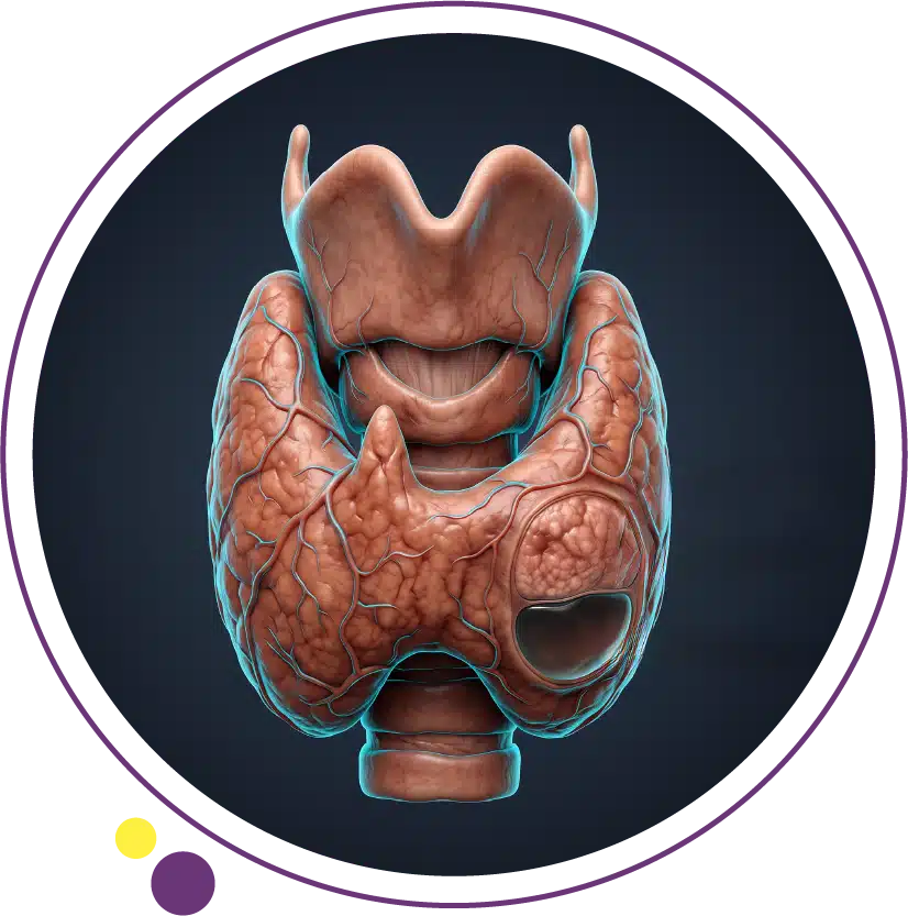
Thyroid nodules are one of the most common incidental findings in modern medical imaging. A patient scheduled for a neck ultrasound for an entirely unrelated reason, or even a CT scan of the chest performed for a completely different concern, frequently turns up a small nodule within the thyroid gland that nobody suspected was there. The vast majority of these findings are harmless, but understanding what they are, how they are classified, and what actually determines whether one needs further investigation helps put an unexpected finding into proper perspective.
In this article, we explain what thyroid nodules are and why they become more common with age, the key difference between solid and cystic nodules, the specific imaging features that determine risk level, when biopsy becomes necessary, and what happens after a nodule is found on a scan.
What Are Thyroid Nodules?
A thyroid nodule is simply a distinct lump or growth of tissue within the thyroid gland that differs from the surrounding normal tissue. Nodules can be a single isolated growth or multiple nodules scattered throughout the gland, and they range enormously in size, from a few millimetres detected only by sensitive imaging to several centimetres large enough to be felt or even seen as a visible neck swelling.
Thyroid nodules are extraordinarily common. Population studies using high-resolution ultrasound have found nodules in a substantial proportion of adults, with prevalence rising steadily with age, and the discovery is frequently incidental, meaning the nodule was found while imaging the neck for an entirely separate reason. The overwhelming majority, well over 90 percent, are benign and require nothing more than periodic monitoring. Images Diagnostic Center in Kuwait provides the ultrasound expertise needed to characterise these findings accurately and determine which small minority genuinely warrant further evaluation.
Why Thyroid Nodules Become More Common With Age
The prevalence of thyroid nodules climbs steadily throughout adult life, and several factors explain this trend. Thyroid tissue, like tissue throughout the body, accumulates small structural changes over decades of normal cellular turnover, and some of these changes manifest as localised areas of glandular overgrowth, cyst formation, or benign tumour development. Hormonal fluctuations across a woman’s reproductive lifespan also contribute to the notably higher prevalence of nodules in women compared to men.
Additionally, the sensitivity of modern ultrasound has increased dramatically over the past few decades, meaning nodules that would have gone entirely undetected in earlier eras are now readily visible. Autopsy studies conducted before modern imaging existed found thyroid nodules in a striking proportion of individuals who had no clinical suspicion of thyroid disease during life, confirming that most nodules are simply a normal feature of an ageing thyroid gland rather than a sign of disease. This context matters because it means an incidentally discovered nodule, particularly in an older adult, is statistically far more likely to represent this normal ageing process than any form of malignancy.
Solid Versus Cystic Nodules: A Fundamental Distinction
One of the first and most important distinctions ultrasound makes when characterising a thyroid nodule is whether it is solid, cystic, or a mixture of both. A purely cystic nodule is essentially a fluid-filled sac within the thyroid tissue, appearing on ultrasound as a well-defined dark space with no internal blood flow. Purely cystic nodules carry an extremely low risk of malignancy, often cited as under 1 percent, and in most cases require only routine monitoring rather than biopsy.
A solid nodule, by contrast, is composed of actual thyroid tissue rather than fluid, and this is the category where the specific ultrasound characteristics described below become clinically important in determining risk. Many nodules fall somewhere in between, described as complex or mixed cystic-solid nodules, containing both fluid components and solid tissue components. In these mixed nodules, it is specifically the solid portion that receives detailed characterisation, since this is where any malignant potential would arise. Ultrasound at Images provides the resolution needed to make this fundamental distinction accurately for every nodule identified.
What Size Actually Matters
A common misconception is that nodule size alone determines malignancy risk, with larger nodules assumed to be automatically more concerning. In reality, size functions differently: it determines whether a nodule meets the threshold for biopsy consideration based on its other ultrasound characteristics, rather than being an independent risk factor on its own. A small nodule with highly suspicious features may warrant biopsy sooner than a considerably larger nodule with entirely reassuring features.
Standardised risk stratification systems, most notably the ACR TI-RADS system, combine several ultrasound features, including composition, echogenicity, shape, margin, and the presence of calcifications, into a points-based score. This score, together with nodule size, determines whether biopsy is recommended, whether follow-up ultrasound is sufficient, or whether no further action is needed at all. The American College of Radiology developed this system specifically to standardise how radiologists communicate risk and to reduce unnecessary biopsies of nodules that carry minimal genuine risk despite their size. This size-and-features combination is precisely why a detailed, systematic ultrasound report matters more than a single measurement in millimetres.
Ultrasound Features That Raise Suspicion
Several specific sonographic characteristics are known to correlate with higher malignancy risk when present in a solid nodule. Hypoechogenicity, meaning the nodule appears darker than surrounding normal thyroid tissue, is more concerning than a nodule that is equally or more echogenic than the surrounding gland. Irregular or poorly defined margins, rather than smooth well-circumscribed borders, raise suspicion because malignant growth tends to invade surrounding tissue in an irregular pattern rather than displacing it cleanly.
A taller-than-wide shape, where the nodule’s vertical dimension exceeds its horizontal dimension on a transverse image, is a specific and fairly reliable indicator of increased risk, thought to reflect a growth pattern that crosses normal tissue planes rather than growing along them. Microcalcifications, tiny punctate bright spots within the nodule, are one of the more established suspicious features and correlate with a specific type of thyroid cancer. Extrathyroidal extension, where a nodule appears to breach the capsule surrounding the thyroid gland, is a more advanced finding that significantly raises concern. Validation studies of these individual features, and of composite scoring systems that combine them, consistently show that the more of these features present together, the higher the probability of malignancy. The ultrasound service at Images systematically documents each of these features for every nodule assessed.
When Biopsy Becomes Necessary
Fine needle aspiration biopsy is recommended when a nodule’s size and ultrasound risk category, taken together, cross an established threshold. This is not an arbitrary decision but follows structured guidelines: a highly suspicious nodule may warrant biopsy at a size as small as one centimetre, while a nodule with entirely benign-appearing features might not warrant biopsy until it reaches a considerably larger size, or may never require biopsy at all regardless of size if it remains purely cystic or shows entirely reassuring characteristics.
The biopsy itself is a straightforward outpatient procedure performed under ultrasound guidance, using a thin needle to withdraw a small sample of cells for cytological examination under a microscope. It requires no incision, no general anaesthesia, and patients typically resume normal activity the same day. The vast majority of biopsy results return benign, providing reassurance and typically leading to a simple follow-up ultrasound schedule rather than any further intervention. When results are indeterminate or suspicious, additional molecular testing or surgical consultation may follow, guided by the specific cytology findings.
What Happens After a Nodule Is Found
When a nodule is identified, whether incidentally or because it was felt or suspected clinically, the pathway typically begins with a dedicated thyroid ultrasound to characterise it fully, alongside blood tests to check thyroid function. A nodule discovered in the context of an overactive thyroid may prompt additional nuclear medicine imaging to determine whether the nodule itself is producing excess hormone, a category generally considered very low risk for malignancy regardless of its ultrasound appearance. For readers who want to understand exactly how a thyroid ultrasound examination works and what to expect step by step, our companion article covers this process in detail.
For nodules that fall below the biopsy threshold, follow-up ultrasound at defined intervals, often six months to two years depending on the initial risk category, monitors for any growth or change in characteristics that would change the management plan. For nodules that meet biopsy criteria, fine needle aspiration provides the tissue diagnosis that determines whether monitoring, surgical referral, or another pathway is appropriate. Throughout this process, imaging remains the central tool guiding every decision. Our broader guide on thyroid disorders provides additional context on how nodules relate to the gland’s overall function and structure.
Frequently Asked Questions
Are most thyroid nodules cancerous?
No. The large majority of thyroid nodules, generally cited at over 90 percent, are entirely benign. Malignancy is the exception rather than the rule, which is exactly why a structured evaluation using ultrasound features and size thresholds exists: to identify the small minority that genuinely warrant biopsy while avoiding unnecessary procedures on the many nodules that pose no real risk.
Can a thyroid nodule disappear on its own?
Purely cystic nodules can shrink or occasionally resolve, particularly if fluid is reabsorbed over time. Solid nodules generally persist and may grow slowly or remain stable in size over years, but true spontaneous resolution of a solid nodule is uncommon. This is one reason follow-up ultrasound, rather than a single assessment, is often recommended for nodules that do not require immediate biopsy.
Does nodule size alone tell me if I need a biopsy?
No. Size works together with the nodule’s specific ultrasound features to determine the biopsy threshold, following structured risk categories rather than size alone. A small nodule with suspicious features may need biopsy sooner than a larger nodule that appears entirely benign on ultrasound. This is why a detailed sonographic assessment matters more than measurement in isolation.
I felt a lump in my neck. Does that mean it’s serious?
A palpable lump warrants evaluation, but feeling a nodule does not itself indicate malignancy. Many entirely benign nodules are large enough to feel, particularly when they occur near the surface of the gland. The appropriate next step is a dedicated thyroid ultrasound to characterise the finding accurately, at which point risk can be properly assessed based on its actual features rather than on the fact that it can be felt.
Where can I get a thyroid ultrasound in Kuwait?
Images Diagnostic Center provides thyroid ultrasound with structured risk assessment, along with ultrasound-guided fine needle aspiration when indicated, across its Jabriya and Salmiya branches in Kuwait. You can contact Images to arrange an evaluation following a clinical referral or an incidental finding on another scan.
Context Turns an Unexpected Finding Into a Manageable One
Discovering a thyroid nodule, whether felt during a physical exam or spotted incidentally on a scan performed for something else entirely, is a common experience that rarely signals anything serious. What matters is a systematic ultrasound evaluation of the nodule’s specific features, applied against established risk criteria, to determine the small minority of cases that genuinely warrant biopsy or closer monitoring.
Images Diagnostic Center provides thyroid ultrasound and fine needle aspiration biopsy across two Kuwait branches:
To arrange a thyroid ultrasound evaluation, contact Images directly.