
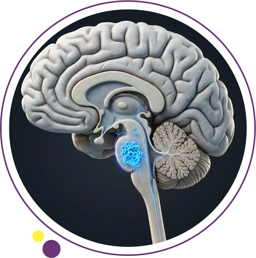
Brain tumor causes are poorly understood compared to many other cancers, and this gap has allowed persistent myths to take hold. Cell phones, head injuries, and power lines are regularly blamed in popular media, but the scientific evidence for most of these concerns is either absent or far weaker than commonly portrayed. Meanwhile, the risk factors that are genuinely established receive far less public attention. This article examines what the evidence actually shows, separates established science from myth, and explains how imaging supports monitoring for those with confirmed risk factors.
Why Brain Tumor Causes Are Difficult to Establish
Primary brain tumors are rare, which limits the statistical power of even large epidemiological studies. Most develop over many years, making it difficult to trace causative exposures from the distant past. Unlike lung cancer where smoking is a dominant measurable cause, brain tumors appear to arise from a complex interplay of genetic susceptibility and environmental factors that differ between tumor types. The only clearly established modifiable environmental risk factor for common primary brain tumors is high-dose ionising radiation. For patients with confirmed risk factors who require monitoring, brain MRI at Images Diagnostic Center in Kuwait provides the sensitivity needed to detect changes at the earliest possible stage.
Ionising Radiation: The One Established Environmental Risk
High-dose therapeutic radiation to the head is the most consistently documented environmental cause of brain tumors. Studies of atomic bomb survivors showed elevated rates of gliomas and meningiomas following high-dose exposure. Patients who received cranial radiation for childhood leukaemia or other head and neck cancers have increased rates of secondary brain tumors appearing five to twenty or more years after original treatment. This finding has driven modern oncology toward radiation-sparing protocols wherever feasible in paediatric treatment.
Long-term survivors of childhood cancers who received cranial radiation should discuss surveillance protocols with their oncologist, which may include periodic brain MRI. Diagnostic radiation doses used in CT and X-ray imaging are far lower than the therapeutic doses associated with elevated risk and are not considered a meaningful brain tumor risk factor. Both CT and MRI services at Images support this surveillance monitoring across three Kuwait branches.
Cell Phones: What the Evidence Actually Shows
Mobile phones emit non-ionising radiofrequency electromagnetic fields that lack sufficient energy to break chemical bonds or damage DNA directly. This is fundamentally different from ionising radiation. The largest studies on this question, including the Interphone study across thirteen countries involving over five thousand brain tumor cases, found no consistent association between phone use and glioma or meningioma. The IARC classified mobile phone radiation as Group 2B, reflecting limited and inconclusive evidence rather than an established causal link.
Critically, brain tumor incidence rates have not increased in the population in the decades since mobile phone use became near-universal, which would be expected if phones were a meaningful cause. The current scientific consensus does not support mobile phone use as an established risk factor. Patients concerned about neurological health should act on symptoms rather than unproven exposures. Our article on brain tumor symptoms describes the warning signs that should prompt evaluation. The imaging services at Images are available to support this clinical assessment in Kuwait.
Head Trauma: Not an Established Cause
Head injury is not an established brain tumor cause despite widespread public belief. Multiple large epidemiological studies examining this relationship have not found consistent causal evidence. Apparent associations in some studies likely reflect surveillance bias: people who receive brain imaging after an injury are more likely to have incidental tumors discovered that would otherwise have gone undetected. There is no established biological mechanism by which mechanical trauma would cause malignant transformation in brain cells.
New neurological symptoms after a head injury warrant evaluation based on the symptoms themselves, not because of brain tumor risk from the injury. CT or MRI at Images can be arranged when clinically indicated based on the symptom picture. The full imaging services at Images support this evaluation across all three Kuwait branches.
Hereditary Syndromes: The Strongest Known Risk Factors
Germline genetic mutations producing hereditary syndromes are the most clearly established and clinically significant brain tumor risk factors. Neurofibromatosis type 1 (NF1) is strongly associated with optic pathway gliomas and astrocytomas. Neurofibromatosis type 2 (NF2) produces bilateral acoustic neuromas and has strong associations with meningiomas. Li-Fraumeni syndrome (TP53 mutations) includes gliomas within its cancer spectrum. Turcot syndrome involves colorectal polyposis combined with glioblastoma or medulloblastoma. Cowden syndrome (PTEN mutations) and constitutional mismatch repair deficiency syndrome also carry elevated brain tumor risk.
Individuals with these hereditary syndromes should be under active neurological and genetic surveillance. Regular brain MRI is a standard component of several syndrome-specific surveillance protocols. The 3 Tesla MRI service at Images provides the resolution needed for detailed neurological monitoring in Kuwait. For patients who need a more comfortable scan environment, Open MRI is also available at Images.
Immune Suppression and CNS Lymphoma Risk
Severe immune suppression significantly increases the risk of primary central nervous system lymphoma. HIV with profound immune deficiency, organ transplant recipients on immunosuppressive therapy, and patients on high-dose immunosuppression for autoimmune conditions all have elevated rates of this condition. The mechanism involves failure of immune surveillance against Epstein-Barr virus-driven B-cell lymphoproliferation within the CNS. Immunocompromised patients who develop new neurological symptoms require brain MRI without delay. The imaging appearance of CNS lymphoma is distinctive and different from glioma, making accurate MRI characterisation clinically important for treatment planning. Brain MRI at Images provides this characterisation with the precision treatment teams rely on.
Age and Gender as Demographic Risk Patterns
Glioblastoma peaks in incidence in the sixth and seventh decades of life. Medulloblastoma and embryonal tumors are predominantly paediatric. Meningiomas are two to three times more common in women, likely reflecting hormonal influences, as meningiomas express progesterone receptors and can grow during pregnancy. These demographic patterns inform clinical assessment but do not support population-level brain tumor screening, since no validated low-risk screening test exists for routine use. Brain MRI for symptomatic evaluation remains the standard clinical approach, and the imaging services at Images are available for this purpose across Kuwait.
What Does Not Cause Brain Tumors
Power lines and extremely low frequency electromagnetic fields have been studied extensively without producing consistent evidence of elevated brain tumor risk. Artificial sweeteners, aluminium exposure, and food additives have not been established as causal factors. Dental X-rays at clinical doses have not been confirmed as meaningful risk factors in well-designed research. Stress and personality factors have no established causal role. The most useful response to concern about brain tumor risk is awareness of the neurological warning signs that should prompt imaging. Acting on genuine symptoms early is far more impactful on outcomes than avoiding unproven exposures. The Images health blog provides evidence-based resources on these topics for patients in Kuwait.
Frequently Asked Questions
Do mobile phones cause brain cancer?
Current scientific consensus does not establish this. The largest studies have not found consistent causal evidence, brain tumor incidence has not risen alongside universal phone adoption, and the IARC Group 2B classification reflects limited rather than established risk. Current evidence does not support restricting phone use for brain cancer prevention.
Can a head injury lead to a brain tumor?
Head injury is not an established brain tumor cause. Apparent associations in some studies reflect surveillance bias. New neurological symptoms after a head injury warrant evaluation based on symptoms, not on the trauma history as a cancer risk factor. MRI at Images can be arranged when clinically indicated.
Are brain tumors hereditary?
Most brain tumors are sporadic rather than inherited. However, specific hereditary syndromes including NF1, NF2, Li-Fraumeni syndrome, and Turcot syndrome carry significantly elevated risk. A family history of multiple brain tumors or a known hereditary syndrome warrants genetic counselling and surveillance imaging at Images.
Should I have a brain MRI due to family history of brain tumors?
A positive family history with multiple affected relatives or a known hereditary syndrome is a reason to discuss surveillance with a neurologist or genetic counsellor. Population-level screening for asymptomatic individuals without a specific syndrome is not currently recommended. For symptomatic patients, brain MRI should be arranged based on clinical presentation. Contact Images to arrange evaluation when clinically indicated.
Evidence Over Anxiety: Focusing on What Genuinely Matters
Understanding actual brain tumor risk factors protects against unnecessary anxiety and focuses attention on what matters most: recognising neurological warning signs early, maintaining surveillance in confirmed hereditary syndrome carriers, and seeking brain MRI promptly when symptoms warrant it. Images Diagnostic Center provides 3 Tesla brain MRI across three Kuwait branches:
To arrange brain imaging in Kuwait, contact Images directly.