
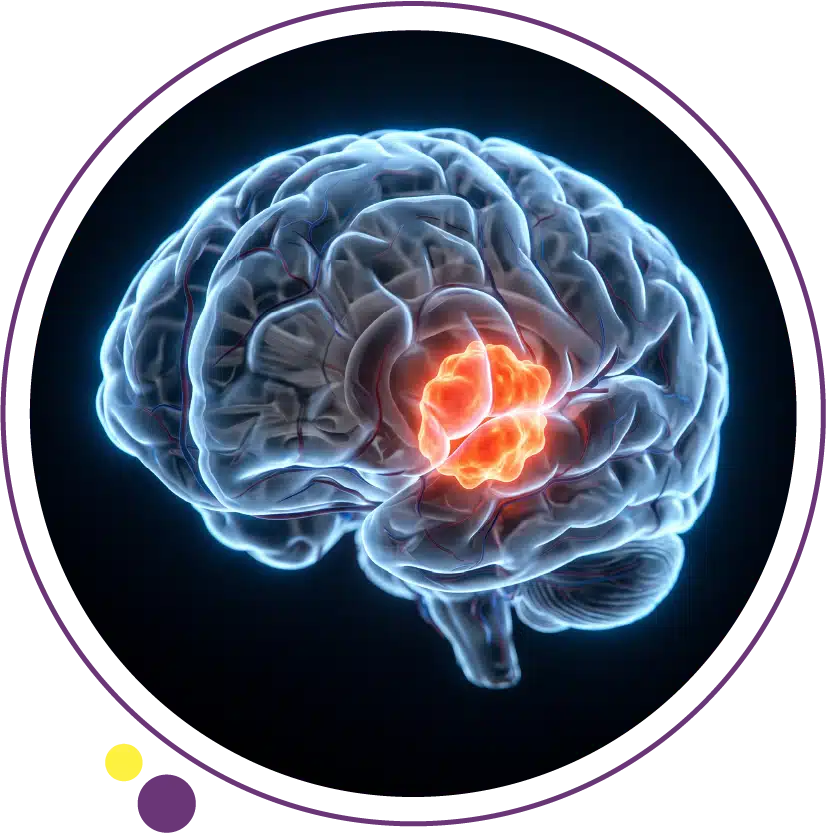
Brain tumor symptoms are among the most variable and locationdependent in all of medicine. A tumor the size of a grape in one part of the brain may produce dramatic neurological deficits immediately recognisable as serious, while the same size tumor in a different region may cause nothing more than a subtle change in personality or the occasional headache that the patient barely notices. This dependence on anatomical location is what makes brain tumor symptoms so clinically challenging and why understanding the relationship between brain structure and function is essential to recognising when a symptom pattern warrants urgent neurological assessment and brain imaging.
In this article, we will explain the eight most common brain tumor symptoms in clinical practice, describe how each symptom arises and what makes it distinctive from more common benign causes, map symptoms to their most likely anatomical locations within the brain, discuss how symptom patterns differ by tumor type, and clarify when brain tumor symptoms constitute a neurological emergency requiring immediate imaging.
Why Brain Tumor Symptoms Depend So Critically on Location
The brain is a highly specialised organ with different regions controlling distinctly different functions. The frontal lobes govern personality, executive function, voluntary movement, and language production. The temporal lobes process hearing, language comprehension, and memory. The parietal lobes integrate sensory information, spatial awareness, and reading. The occipital lobes are devoted almost entirely to vision processing. The cerebellum coordinates movement, balance, and fine motor control. The brainstem controls fundamental autonomic functions including breathing, heart rate, and consciousness, as well as serving as the conduit for all signals travelling between the brain and the body.
A tumor growing in any of these regions will produce deficits in the functions controlled by that region. This neurological localisation is one of the most powerful tools in clinical neurology: a skilled clinician can often determine from the pattern of symptoms where in the brain a lesion is likely to be located before any imaging is performed. Brain MRI is then used to confirm the clinical suspicion, characterise the lesion in detail, and guide treatment planning. At Images Diagnostic Center in Kuwait, 3 Tesla MRI provides the field strength and resolution needed for this demanding neurological evaluation.
The 8 Most Common Brain Tumor Symptoms
1. Headaches
Headache is one of the most common symptoms in human medicine, and the vast majority of headaches are caused by tension, migraine, or cluster headache patterns that are benign. However, brain tumor headaches have several distinguishing characteristics that should raise clinical concern. The classic brain tumor headache is worse in the morning upon waking, improves as the day progresses, is worsened by the Valsalva manoeuvre (bearing down, coughing, or straining), and is associated with nausea or vomiting. This pattern reflects the position-dependent increase in intracranial pressure that occurs when the patient is lying flat during sleep.
Other headache features that warrant neurological evaluation include a new headache in a patient over fifty without a prior headache history, a headache described as the worst of the patient’s life (thunderclap), a progressive worsening headache that does not respond to standard analgesia, a headache associated with any new neurological symptom including vision change, speech difficulty, or limb weakness, and a headache in a patient with a known systemic cancer where brain metastases are possible. Headache in brain tumors occurs in approximately fifty percent of patients but is typically not the sole presenting symptom. Brain MRI with contrast is the definitive investigation when these red flag features are present.
2. Seizures
A new-onset seizure in an adult who has never had a seizure before is a neurological emergency and requires brain imaging to exclude a structural cause. Brain tumors are among the most important structural causes of new-onset adult seizures, particularly for tumors located in or near the cortex where they irritate the neuronal circuitry and lower the seizure threshold. Tumors in the frontal and temporal lobes most commonly present with seizures, reflecting the proximity of these regions to seizure-generating cortical circuits.
Seizures from brain tumors can take many forms: generalised tonic-clonic convulsions, focal motor seizures affecting one limb or one side of the face, focal sensory seizures producing tingling or visual disturbances, or complex partial seizures causing altered awareness with automatic behaviours. Any new seizure in an adult should be followed by brain MRI with contrast as a priority, ideally within twenty-four to forty-eight hours. The CT scan is often used as the emergency study when MRI is not immediately available, but MRI subsequently provides far superior characterisation of any lesion identified. At Images in Kuwait, brain MRI is available across all three branches for this urgent clinical need.
3. Vision Changes
Vision disturbances from brain tumors reflect either direct involvement of the visual pathways or raised intracranial pressure affecting the optic nerves. Papilloedema, which is swelling of the optic disc visible on fundoscopic examination, develops when raised intracranial pressure is transmitted to the optic nerve sheath and is one of the most important clinical signs of raised pressure within the skull. Patients may describe blurred vision, transient visual obscurations lasting seconds when they change posture, or a reduction in peripheral vision.
Specific patterns of visual field loss help localise the tumor site. A tumor compressing the optic chiasm, which is where the two optic nerves cross, produces a characteristic bitemporal hemianopia: loss of the outer half of the visual field in both eyes. This is the classic visual presentation of a pituitary tumor. A tumor in the occipital lobe produces contralateral homonymous hemianopia: loss of the same half of the visual field in both eyes. Double vision (diplopia) may result from cranial nerve compression along the base of the skull or within the brainstem, and is a sign that warrants urgent imaging. Brain MRI with gadolinium contrast at Images provides the detail needed to identify these cranial nerve and optic pathway lesions with precision.
4. Speech and Language Problems
Tumors in or near the language areas of the dominant hemisphere (almost always the left hemisphere in right-handed individuals and in most left-handed individuals) produce speech and language deficits that are clinically specific and highly localising. Broca’s area in the posterior inferior frontal lobe is responsible for speech production. Damage here produces expressive aphasia: the patient understands what is said to them but has difficulty finding words and producing fluent speech. Wernicke’s area in the superior temporal lobe is responsible for language comprehension. Damage here produces receptive aphasia: fluent but meaningless speech with profound difficulty understanding spoken or written language.
New-onset speech difficulties in an adult who had normal speech previously, particularly if progressive over weeks, should always prompt neurological evaluation and brain imaging. The MRI service at Images provides the high-resolution cortical imaging needed to identify and characterise tumors in these language-critical brain regions. For patients who may benefit from understanding the broader spectrum of neurological symptoms that indicate a need for brain MRI, including those related to demyelinating disease, our previously published articles on multiple sclerosis symptoms and MS diagnosis provide useful comparative context on neurological symptom evaluation.
5. Memory and Cognitive Changes
Cognitive changes including memory impairment, difficulty with concentration, slowed thinking, or a general decline in intellectual performance can reflect tumor involvement of the frontal or temporal lobes, or the widespread effects of raised intracranial pressure on global brain function. These symptoms are among the most difficult to recognise as neurological in origin because they can appear gradually and are frequently attributed to stress, sleep deprivation, aging, or depression by both the patient and their primary care physician.
The clinically significant feature that distinguishes tumor-related cognitive change from normal aging or functional cognitive complaint is the progressive nature of the deterioration. A person who was previously functioning normally cognitively and is now significantly impaired within weeks to months of symptom onset, particularly in the context of any other neurological symptom, has a pattern that demands imaging. Neuropsychological testing can document and quantify the cognitive impairment, but brain MRI with contrast at Images determines the structural cause and guides the further assessment and management pathway.
6. Weakness or Numbness in Limbs
Unilateral limb weakness or numbness, affecting one arm, one leg, or one entire side of the body, reflects disruption of the motor or sensory pathways descending from or ascending to the contralateral cerebral hemisphere. The motor cortex runs in a strip along the posterior frontal lobe, with different body regions represented in a topographic map: the leg and foot medially, the arm and hand laterally, and the face at the lowest point. A tumor in the motor cortex produces a contralateral upper motor neuron weakness pattern: increased tone, brisk reflexes, and impaired voluntary movement.
Progressive unilateral limb weakness that has developed over weeks or months and is not explained by a peripheral nerve or spinal cause is a significant neurological finding requiring brain MRI. The onset of sudden severe limb weakness, particularly in the context of headache or altered consciousness, may indicate acute hemorrhage into a brain tumor, which is a neurological emergency. At Images, brain MRI and CT are both available to support emergency evaluation of these presentations, with CT being the faster study when acute haemorrhage is the primary concern.
7. Balance and Coordination Problems
Cerebellar tumors and posterior fossa tumors produce a characteristic constellation of symptoms related to the cerebellum’s role in coordinating movement and maintaining balance. Gait ataxia, which is an unsteady wide-based walking pattern, limb ataxia affecting the precision of arm and hand movements, nystagmus (involuntary rhythmic eye movements), dysarthria (slurred scanning speech), and dizziness or vertigo of a central character are all signs of cerebellar dysfunction. Posterior fossa tumors including medulloblastoma in children and haemangioblastoma or metastatic tumors in adults are among the diagnoses in this anatomical region.
Balance problems arising from brain tumors are distinct from the benign positional vertigo caused by inner ear conditions (labyrinthine pathology) in several ways: cerebellar ataxia is not typically position-dependent in the same stereotyped way as benign positional vertigo, it may be accompanied by other neurological signs, and it does not respond to the repositioning manoeuvres that are effective for inner ear causes. Any unexplained progressive balance problem or ataxia that is not explained by inner ear disease should prompt brain MRI. The high-resolution posterior fossa imaging provided by 3 Tesla MRI at Images is particularly well suited to evaluating this region, which can be technically challenging at lower field strengths. For patients anxious about enclosed MRI scanners, Open MRI is also available at Images.
8. Personality and Behavior Changes
Personality and behaviour changes are among the most difficult brain tumor symptoms to recognise because they may be subtle, gradual, and initially attributed to psychiatric illness or life circumstances. A previously organised and punctual person who becomes disinhibited, impulsive, or apathetic; a patient whose social judgement has deteriorated; someone who has become irritable or emotionally flat without apparent reason; or a person whose motivation and initiative have declined significantly may all be exhibiting the frontal lobe signs of a tumor in this region. Frontal lobe tumors, including gliomas and meningiomas in the frontal region, are particularly associated with these personality changes because of the frontal lobe’s role in executive function and social behaviour.
These changes are often first noticed by family members or close colleagues rather than by the patient, who may lack insight into their own changed behaviour due to the brain region affected. A psychiatric referral without a prior brain MRI to exclude a structural cause in a patient with new-onset behavioural or personality change is an incomplete assessment. New-onset psychiatric-like symptoms in middle-aged or older adults with no prior psychiatric history should routinely include brain MRI. The full imaging services at Images Diagnostic Center support the neurological evaluation of these presentations in Kuwait.
How Symptoms Map to Brain Locations
Frontal lobe tumors produce personality change, executive dysfunction, impaired planning and organisation, contralateral limb weakness (particularly the motor strip), and expressive language difficulty when the dominant hemisphere is involved. Temporal lobe tumors produce seizures, memory impairment, receptive aphasia when the dominant hemisphere is involved, and hearing or smell changes. Parietal lobe tumors produce contralateral sensory loss, difficulty with spatial awareness and navigation, neglect of one side of space when the non-dominant hemisphere is involved, and reading or calculation difficulties when the dominant hemisphere is affected.
Occipital lobe tumors primarily produce visual symptoms including contralateral visual field loss. Cerebellar tumors produce ataxia, dysarthria, and nystagmus. Brainstem tumors produce multiple cranial nerve palsies, crossed signs (ipsilateral face and contralateral limb symptoms reflecting the anatomy of the brainstem), and signs of long tract involvement including limb weakness and sensory loss. Pituitary region tumors produce bitemporal visual field loss and endocrine disturbances. This anatomical symptom mapping is the reason that brain MRI with dedicated sequences at Images is so important: the image needs to answer the specific clinical question raised by the symptom pattern rather than providing a generic brain overview.
When Brain Tumor Symptoms Are a Neurological Emergency
Certain brain tumor presentations require emergency assessment rather than an outpatient neurology appointment. Sudden severe headache described as the worst of the patient’s life suggests haemorrhage, which can occur into a previously asymptomatic tumor. Rapid onset of limb weakness or speech difficulty over minutes to hours may reflect hemorrhage or acute brain herniation secondary to rising intracranial pressure around a large tumor. Reduced level of consciousness in a patient with known or suspected brain tumor is a medical emergency indicating severe intracranial hypertension or herniation. Prolonged or recurrent seizures from a brain tumor also require emergency management.
For non-emergency but urgent presentations including a new seizure, progressive headache with neurological features, or progressive focal neurological deficit, brain MRI should be arranged within twenty-four to forty-eight hours. CT brain is used as the emergency study when MRI is not immediately available, principally to exclude haemorrhage and mass effect requiring emergency neurosurgical intervention. For all subsequent characterisation and treatment planning, MRI provides far superior information. The CT and MRI services at Images are equipped to support both emergency and urgent brain tumor evaluation in Kuwait across the Jabriya, Hawally, and Salmiya branches.
The Critical Role of MRI in Evaluating Brain Tumor Symptoms
MRI is the imaging modality of choice for brain tumor evaluation. Its superior soft tissue contrast compared to CT allows exquisite visualisation of the brain parenchyma, identification of subtle infiltrative tumors, characterisation of tumor boundaries, assessment of peritumoral oedema, and detection of leptomeningeal disease. Contrast-enhanced MRI identifies the blood-brain barrier breakdown characteristic of high-grade tumors, with the enhancement pattern providing diagnostic information about tumor grade and type. Specific MRI sequences including FLAIR (fluid-attenuated inversion recovery), diffusion-weighted imaging, and MR spectroscopy add further characterisation that helps distinguish different tumor types and grades.
The 3 Tesla MRI at Images provides the field strength that maximises signal-to-noise ratio in brain imaging, enabling detection of smaller lesions and more detailed characterisation of tumor architecture than lower-field systems. This level of detail is directly relevant to clinical outcomes because it influences the accuracy of treatment planning. For patients who have difficulty tolerating enclosed scanners, the Open MRI at Images provides a more comfortable alternative that can be used for most brain evaluations. The Images health blog contains additional patient-focused resources on MRI procedures, what to expect, and how to prepare.
Frequently Asked Questions
Can a brain tumor cause only headaches without other symptoms?
Yes, in early stages a brain tumor can produce only headaches before other neurological deficits appear. However, a headache as the sole brain tumor symptom is relatively uncommon, and when it is the presenting complaint, the headache typically has the features described above: progressive, worse in the morning, worsened by Valsalva manoeuvre, and associated with nausea. Most patients with brain tumors have at least one additional symptom alongside headache. Any headache meeting the red flag criteria should prompt brain MRI regardless of whether other symptoms are present.
How quickly do brain tumor symptoms develop?
The rate of symptom development depends strongly on tumor type and grade. High-grade glioblastoma can progress from minimal symptoms to severe disability within weeks because of its rapid growth and extensive surrounding oedema. Low-grade gliomas may grow so slowly that symptoms develop imperceptibly over years, with patients noticing only subtle changes retrospectively. Meningiomas, which are typically benign, may be present for many years before producing noticeable symptoms because they grow slowly and displace rather than invade brain tissue. The rate of symptom progression is itself a diagnostic clue that MRI can correlate with tumor characteristics.
Is CT or MRI better for evaluating brain tumor symptoms?
MRI is substantially superior to CT for brain tumor evaluation in most circumstances. CT is faster and more widely available for emergency situations, and is the first-line study when haemorrhage needs to be excluded rapidly. However, CT misses many smaller tumors, infiltrative gliomas, leptomeningeal disease, and posterior fossa lesions due to bone artefact in the posterior cranial fossa. Once the clinical situation is stable enough to allow MRI, it should always be performed to fully characterise the abnormality. MRI at Images with contrast provides the diagnostic detail that neurosurgical and neuro-oncology teams need for treatment planning.
Can brain tumor symptoms mimic other conditions?
Yes, frequently. Headaches are most commonly benign. Personality changes are often attributed to depression or anxiety. Memory difficulties are attributed to aging or stress. Seizures may initially be misclassified. Balance problems may be attributed to inner ear disease. Vision changes may prompt ophthalmological evaluation without neurological referral. This is precisely why a comprehensive brain MRI is the appropriate investigation whenever any of these symptoms has a neurological character or occurs in the clinical context described in this article, rather than waiting for the full symptom constellation to develop.
Where can I arrange a brain MRI in Kuwait?
Images Diagnostic Center provides 3 Tesla brain MRI with and without contrast across three Kuwait branches, supported by expert radiology reporting. Brain MRI for symptoms including headache, seizure, vision change, speech difficulty, limb weakness, balance problems, cognitive change, and personality change can be arranged by contacting the Images team directly once a clinical referral has been made. Open MRI is also available for patients who require a more comfortable scan environment.
Recognising Brain Tumor Symptoms Before They Progress
The eight symptoms described in this article span a spectrum from the familiar (headache) to the unfamiliar (personality change). What they share is that each one, in the right clinical context and with the right pattern of development, indicates that brain imaging is needed rather than watchful waiting or symptomatic treatment alone. The neurological architecture of the brain is precise enough that symptoms often point toward a specific region, and a brain MRI with contrast will confirm or refute the clinical suspicion with a level of detail that no other investigation can match.
Images Diagnostic Center provides 3 Tesla MRI across three Kuwait branches to support the full evaluation of brain tumor symptoms from initial clinical suspicion through to pre-operative planning:
To arrange brain MRI for neurological symptom evaluation, contact Images directly.