
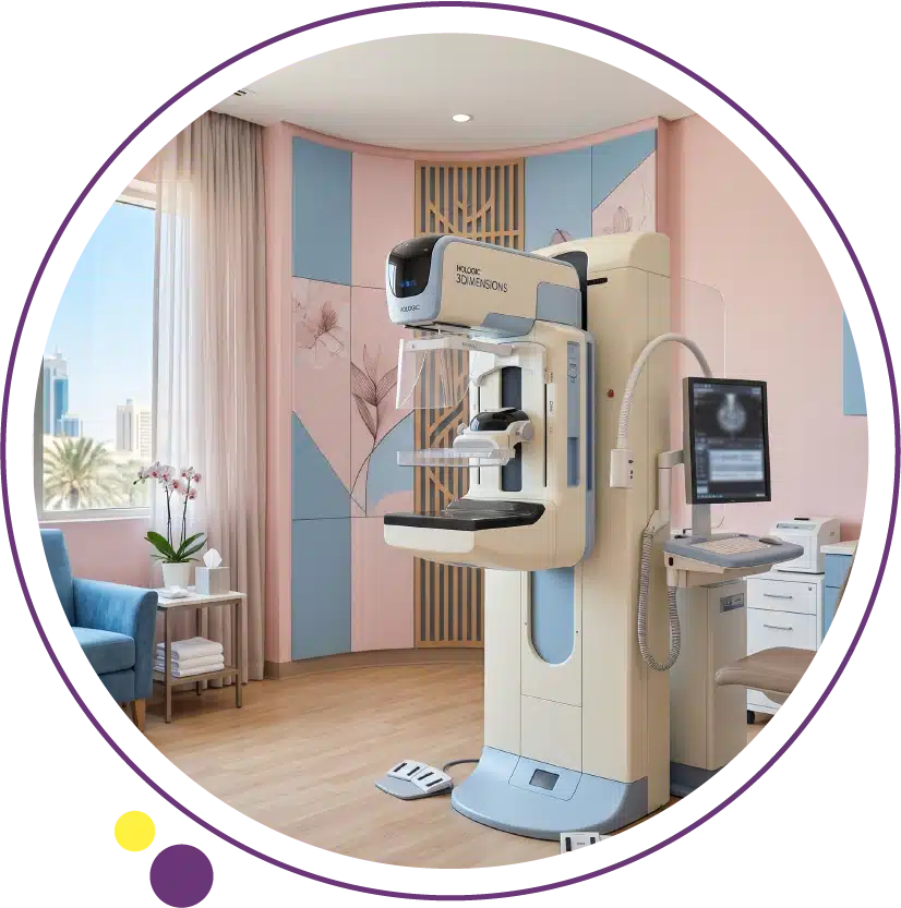
Breast cancer screening is not a single test with a single protocol. It is a family of imaging and clinical tools that are applied in different combinations and at different intervals depending on a woman’s individual risk profile. For the majority of women in the average-risk category, annual or biennial mammography is the standard recommendation. For women with significantly elevated lifetime risk due to genetic mutations, strong family history, or prior chest radiation, the approach is more intensive and begins at a younger age.
Understanding what each screening option can detect, what its limitations are, how the options complement each other, and which schedule applies to which risk group empowers women to make informed decisions and ensures that no opportunity for early detection is missed. In this article, we will compare all the major breast cancer screening options, explain how risk stratification changes the approach, address the specific challenge of dense breast tissue, and describe what happens when a screening study identifies something that requires further evaluation.
Why Breast Cancer Screening Matters
Breast cancer detected at an early stage, before it has spread to regional lymph nodes or distant organs, carries a fundamentally different prognosis from the same cancer detected after metastatic spread. Five-year survival for localised breast cancer exceeds ninety-five percent with modern treatment. Five-year survival for metastatic breast cancer is below thirty percent. The purpose of screening is to bridge this gap by identifying cancers at a stage when curative treatment is the most probable outcome, rather than waiting for symptoms to develop when the disease may already be more advanced.
Screening mammography has been shown in multiple large clinical trials to reduce breast cancer mortality by approximately twenty to forty percent in the screened population compared to unscreened controls, and this benefit is most pronounced when screening is maintained consistently over time. The addition of MRI for high-risk women and ultrasound for women with dense breasts further improves detection rates in specific populations. At Images Diagnostic Center in Kuwait, mammography, ultrasound, and MRI are available to support the full spectrum of breast cancer screening needs.
Mammography: The Standard Screening Foundation
Standard Digital Mammography
Digital mammography uses low-dose X-rays to produce images of the internal breast tissue in two projections: craniocaudal (from above) and mediolateral oblique (from the side). It detects masses, architectural distortion, asymmetries, and calcification patterns that may indicate malignancy, often before a cancer is large enough to be felt by a woman or her clinician. The entire examination takes approximately fifteen to twenty minutes and involves brief compression of each breast between two plates to obtain clear, detailed images.
Mammography performs best in women with fatty or mixed density breast tissue, where tumors stand out as denser areas against a lighter background. Its sensitivity is lower in women with very dense breast tissue, where the dense glandular tissue and a potential tumor are of similar density and the tumor may not be visible. Even with this limitation, mammography remains the recommended primary screening tool for all average-risk women because of its established mortality benefit, accessibility, relatively low radiation dose, and decades of supporting evidence. The mammography service at Images provides high-quality digital imaging with expert reporting for patients in Kuwait.
3D Mammography (Digital Breast Tomosynthesis)
Digital breast tomosynthesis, commonly called 3D mammography, acquires multiple low-dose X-ray images at different angles and reconstructs them into a series of thin cross-sectional images through the breast tissue. This resolves one of the key limitations of standard 2D mammography: the overlapping of breast tissue layers, which can either obscure a real tumor or create false-positive-appearing densities that result in unnecessary recall and biopsy. Multiple studies have shown that 3D mammography increases cancer detection rates and reduces recall rates compared to standard 2D mammography, with particularly meaningful benefits in women with dense breasts.
The total radiation dose from a combined 2D and 3D examination is approximately twice that of 2D alone, though still well within the range considered safe for annual use. Increasingly, tomosynthesis is offered as the primary mammography option at breast centres that have adopted the technology. Women with dense breasts, prior indeterminate mammography findings, or high-risk features who are having their annual mammography may find that 3D mammography offers a meaningful improvement in detection sensitivity. The imaging team at Images can advise which mammography modality is most appropriate for individual patients based on their clinical history.
Ultrasound: A Valuable Supplement for Dense Breasts
Breast ultrasound uses high-frequency sound waves to produce real-time images of breast tissue and does not involve ionising radiation. Its primary role in breast cancer screening is as a supplement to mammography in women with dense breasts, where it can detect mammographically occult cancers that are hidden within the dense fibroglandular tissue. Studies combining mammography with ultrasound in women with dense breasts have consistently found additional cancers beyond those detected by mammography alone, at a rate of approximately three to four additional cancers per thousand women screened.
Ultrasound is also the standard next step when mammography identifies a mass that requires characterisation. The distinction between a simple fluid-filled cyst (benign) and a solid mass that requires biopsy is made definitively by ultrasound in most cases. Breast ultrasound at Images supports both of these clinical functions: supplemental screening for dense breast tissue and characterisation of mammographic findings. One limitation of ultrasound as a screening tool is its operator-dependence and the higher rate of false-positive recalls compared to mammography, which is why it is generally used as a supplement rather than a replacement. For a comprehensive overview of what a breast cancer workup involves after detection, our article on breast cancer diagnosis covers the full diagnostic pathway.
Breast MRI: The Most Sensitive Tool for High-Risk Women
Contrast-enhanced MRI of the breast is the most sensitive imaging modality for breast cancer detection, with sensitivity exceeding eighty to ninety percent compared to approximately seventy to eighty percent for mammography in dense breasts. It detects cancers that are invisible on both mammography and ultrasound, and is particularly valuable for identifying multifocal or multicentric disease and contralateral cancers that would be missed by breast-only evaluation. These properties make it the preferred additional screening tool for women at significantly elevated lifetime risk.
The main limitations of breast MRI in a screening context are its higher false-positive rate compared to mammography, which leads to more recalls and benign biopsies, its requirement for intravenous contrast injection, its longer examination duration, and its higher cost. These limitations make it impractical and unjustifiable for universal population-level screening but fully warranted for high-risk women where the pre-test probability of malignancy is substantially higher. Guidelines from major radiology and oncology societies specify which risk categories qualify for annual breast MRI screening. At Images, 3 Tesla MRI provides the field strength and spatial resolution needed for the demanding diagnostic requirements of breast MRI screening.
Risk Stratification: How Screening Differs by Risk Level
Average-Risk Women
Average-risk women are those without a personal or strong family history of breast cancer, without known BRCA1 or BRCA2 mutations, without a history of prior chest radiation at a young age, and without other high-risk conditions. For this group, the standard recommendation from most major guidelines is annual mammography beginning at age forty to fifty, with specific starting age recommendations varying between guidelines. The American Cancer Society recommends the option to begin at forty with annual screening, and mandates it from forty-five. Many European guidelines recommend biennial screening from fifty. The choice between annual and biennial, and between starting at forty or fifty, is a clinical decision best made in consultation with a physician who takes into account individual preferences and the local guideline framework.
Average-risk women do not typically require supplemental ultrasound or MRI screening unless dense breast tissue is identified on mammography, at which point a discussion about supplemental imaging is warranted. For women in this category, annual mammography at Images in Kuwait provides the evidence-based standard of screening care. For a comprehensive discussion of the signs of breast cancer that may arise between screening rounds, our article on signs of breast cancer provides a complete clinical guide.
Intermediate-Risk Women
Women with a lifetime breast cancer risk of fifteen to twenty percent, often estimated through formal risk models such as the Tyrer-Cuzick or Claus models, are classified as intermediate risk. This group may include women with a family history of breast cancer that does not meet criteria for hereditary syndrome testing, women with prior atypical ductal hyperplasia or lobular carcinoma in situ, and women with dense breast tissue on mammography. For intermediate-risk women, annual mammography beginning at forty is generally recommended, with the possible addition of annual ultrasound when breast density is classified as heterogeneously or extremely dense. The specific supplemental imaging recommendation depends on the clinical assessment.
Intermediate-risk women may be offered MRI in some clinical settings based on their specific combination of risk factors, though the evidence base for this is less established than for the high-risk category. The imaging team at Images can provide the mammography and ultrasound components of a tailored screening programme for these women in Kuwait, with MRI available when clinically indicated by their managing physician.
High-Risk Women
Women with a lifetime breast cancer risk of twenty to twenty-five percent or higher, confirmed BRCA1 or BRCA2 mutations or untested first-degree relatives of BRCA carriers, a prior history of chest radiation between ages ten and thirty, or certain hereditary syndromes including Li-Fraumeni and Cowden syndrome are classified as high risk. For these women, annual breast MRI combined with annual mammography is the recommended standard in most international guidelines, beginning at age twenty-five to thirty depending on the specific risk condition. The two modalities are complementary and detect different subsets of cancers; using both maximises detection sensitivity.
ACR and ACS guidelines for high-risk women represent the clearest and most evidence-supported framework for the supplemental MRI recommendation. Women who fall into the high-risk category and are not yet in an intensive screening programme should discuss initiating one with their physician without delay. The MRI service and mammography service at Images are both available to support annual dual-modality screening for high-risk women in Kuwait.
Dense Breast Tissue: An Important Screening Variable
Breast density is assessed on every mammogram and reported using the ACR BI-RADS density categories: almost entirely fatty (A), scattered fibroglandular density (B), heterogeneously dense (C), or extremely dense (D). Women with categories C or D, who represent approximately forty to fifty percent of all women undergoing mammography, have two related concerns: their mammography sensitivity is lower because tumors are harder to distinguish from dense tissue, and dense tissue itself is an independent risk factor for developing breast cancer.
For women with dense breasts who receive a normal mammogram result, supplemental ultrasound screening can identify additional cancers that mammography missed. Supplemental MRI provides the highest incremental cancer detection rate but comes with the tradeoffs described above. Many institutions and national guidelines now require mammography reports to include notification of breast density so that patients can have an informed conversation with their physician about supplemental screening. When the density result comes back as heterogeneously or extremely dense, a discussion with the Images team about supplemental ultrasound is a practical next step.
What Happens When Screening Finds Something
A screening study that identifies a finding requiring further evaluation is not a cancer diagnosis. The vast majority of findings recalled for additional assessment after mammography turn out to be benign. The recall process involves returning for additional imaging, typically spot compression mammography views to better characterise an asymmetry, or ultrasound to evaluate a mass identified on mammography. If the additional imaging is reassuring, the patient is returned to routine screening. If the finding is suspicious, biopsy is recommended.
Biopsy for breast lesions is typically performed using a minimally invasive ultrasound-guided or stereotactic (mammography-guided) core needle technique. This provides histopathological tissue without the need for open surgical biopsy in most cases. If biopsy confirms malignancy, staging workup and treatment planning begin. If biopsy returns benign results, the patient is returned to screening with appropriate surveillance imaging to confirm stability of the benign lesion. The ultrasound and mammography services at Images support the diagnostic assessment step for patients whose screening identifies a finding requiring further evaluation in Kuwait.
Frequently Asked Questions
At what age should breast cancer screening begin?
For average-risk women, most guidelines recommend beginning annual mammography at age forty to fifty. For high-risk women including BRCA mutation carriers and those with equivalent high-risk conditions, annual mammography plus MRI typically begins at twenty-five to thirty. For women with intermediate risk factors, the decision is individualised by their physician. The starting age should be discussed with a doctor who can take into account personal and family history, breast density, and any known genetic risk factors.
Is breast MRI better than mammography for screening?
MRI is more sensitive than mammography, meaning it finds more cancers. However, it also has a higher false-positive rate, meaning it more frequently identifies findings that turn out to be benign and require unnecessary biopsy. This higher false-positive rate makes MRI impractical for universal screening but justified for high-risk women where the pre-test probability of cancer is substantially higher. Mammography and MRI are complementary rather than competing: both detect different cancers, and high-risk women benefit most from using both together annually.
Does breast ultrasound replace mammography?
No. Breast ultrasound is a valuable supplement to mammography but does not replace it. Mammography detects calcification patterns that are invisible on ultrasound and that represent a significant proportion of clinically important findings including ductal carcinoma in situ. Ultrasound compensates for mammography’s reduced sensitivity in dense breast tissue by detecting masses that are obscured on mammogram. The two modalities work together most effectively when used in combination rather than one in place of the other.
How often should high-risk women be screened?
Annual mammography and annual breast MRI are the standard recommendation for high-risk women in most guidelines, with the two studies typically alternated at six-month intervals so that some form of breast imaging is performed every six months throughout the year. This intensive approach reflects the higher background cancer rate in this population and the more aggressive biological behaviour of cancers that develop in BRCA mutation carriers. The schedule is tailored by the physician managing the patient’s high-risk surveillance programme.
Is breast cancer screening available in Kuwait?
Yes. Images Diagnostic Center provides mammography, breast ultrasound, and 3 Tesla MRI across three branches in Kuwait, covering the full spectrum of breast cancer screening modalities from standard annual mammography for average-risk women to combined mammography and MRI for high-risk women. Appointments can be arranged by contacting the Images team directly.
Matching the Right Screening to the Right Risk Level
Breast cancer screening is most effective when it is tailored to the individual rather than applied uniformly. A one-size-fits-all approach leaves high-risk women under-screened and exposes average-risk women to unnecessary investigations. Knowing your risk level, understanding which screening modalities apply to that risk level, and maintaining a consistent annual schedule are the three most practically important steps any woman can take to benefit from what screening has to offer. The imaging team at Images can provide all the screening modalities described in this article across Kuwait’s three branches.
To schedule your mammography, ultrasound, or MRI breast screening, contact Images directly.