
Receiving a DEXA scan report can feel confusing if you are not familiar with what the numbers mean. T-scores, Z-scores, and bone mineral density figures in unfamiliar units can leave patients uncertain about whether their results are reassuring, concerning, or somewhere in between. The good news is that DEXA scan interpretation follows a clear and standardised framework that, once understood, makes your results immediately meaningful and actionable.
In this article, we will explain exactly what a DEXA scan measures, how to read the T-score and Z-score, what the different result ranges mean clinically, how the FRAX fracture risk tool uses your results, what factors can influence your bone density score, what steps to take based on your results, and how often DEXA scanning should be repeated to monitor bone health over time.
What Does a DEXA Scan Actually Measure?
A DEXA scan, which stands for Dual-Energy X-ray Absorptiometry, measures bone mineral density (BMD) by passing two low-energy X-ray beams at different energy levels through the body. Because bone absorbs more X-ray energy than soft tissue, and because denser bone absorbs even more than less dense bone, the difference in absorption between the two beams allows the machine to calculate the mineral content per unit area of the scanned bone.
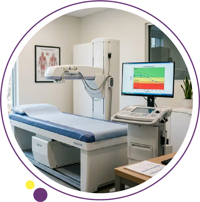
The result is expressed as grams of bone mineral per square centimetre (g/cm²) for each measurement site. Standard DEXA scan sites include the lumbar spine (typically L1 to L4), the femoral neck, and the total proximal femur, as these are the areas most clinically relevant to osteoporosis-related fracture risk. A forearm scan may also be performed in certain situations, such as when hip or spine measurements are unreliable due to structural changes or previous surgery. The raw BMD values are then compared to reference populations to generate the T-score and Z-score that appear prominently on your report. At Images Diagnostic Center in Kuwait, DEXA scanning is available with full reporting across all three branches.
Understanding the T-Score: The Core of DEXA Interpretation
The T-score is the most important number on your DEXA report for assessing osteoporosis risk in adults over fifty. It compares your measured bone mineral density to the mean BMD of a healthy young adult reference population of the same sex, and expresses the difference as a number of standard deviations above or below that reference mean. The World Health Organisation criteria use the T-score to define three clinically distinct categories that determine how bone health is classified and how treatment decisions are made.
Normal Bone Density: T-Score Above -1.0
A T-score of -1.0 or above indicates normal bone mineral density. Your bones are as dense or denser than the average healthy young adult peak bone mass, and your fracture risk from low bone density alone is not elevated. A normal T-score is reassuring but does not mean bone density monitoring is unnecessary in the future. Bone density naturally declines with age, particularly in women after menopause and in men after seventy, so a normal result at one time point does not guarantee that it will remain normal.
If your T-score is normal and you have no major clinical risk factors for osteoporosis, repeat DEXA scanning is typically recommended every two to five years depending on your age and risk profile. Your doctor will advise on the appropriate interval for your situation. The DEXA service at Images makes it straightforward to schedule these repeat assessments at a convenient branch in Kuwait.
Osteopenia: T-Score Between -1.0 and -2.5
A T-score between -1.0 and -2.5 indicates osteopenia, which means your bone density is below the young adult mean but has not yet reached the threshold for an osteoporosis diagnosis. Osteopenia is not a disease in itself; it describes a state of reduced bone density that places you at a modestly higher risk of fracture than someone with normal bone density, while the absolute fracture risk remains lower than for osteoporosis. Approximately 34 percent of postmenopausal women in the United States have osteopenia at the hip.
An osteopenia result does not automatically mean you need medication. Management is individualised and depends on the absolute fracture risk assessment, which takes into account not only the T-score but your age, sex, family history, history of prior fractures, use of glucocorticoid medications, smoking history, and alcohol intake. The FRAX tool, described below, integrates these factors into a ten-year fracture probability that guides treatment decisions. Lifestyle measures including adequate calcium and vitamin D intake, weight-bearing exercise, and fall prevention are recommended for everyone with osteopenia. The medical imaging team at Images can provide your DEXA results with clear reporting that supports your doctor’s clinical assessment.
Osteoporosis: T-Score at or Below -2.5
A T-score of -2.5 or lower confirms a diagnosis of osteoporosis by the WHO criteria. This means your bone mineral density is 2.5 standard deviations or more below the young adult reference mean, representing a significant reduction in bone strength and a substantially elevated risk of fracture from relatively minor trauma, such as a fall from standing height. The most clinically significant fragility fractures occur at the hip, vertebral bodies, and distal forearm. Hip fractures in particular carry significant morbidity and mortality, particularly in older adults, underscoring the importance of treating osteoporosis before a fracture occurs.
An osteoporosis diagnosis on DEXA typically leads to a clinical review of all contributing factors, fracture risk assessment using the FRAX tool, blood tests to exclude secondary causes of bone loss, and a discussion about pharmacological treatment options including bisphosphonates, denosumab, or other agents depending on the patient’s specific situation. It is also worth noting that severe osteoporosis is defined as a T-score of -2.5 or below in the presence of one or more fragility fractures. The DEXA scan result and its clinical interpretation together form the starting point for an effective bone protection plan. For patients who want to understand the broader context of bone imaging, our article on diagnostic X-ray explains how plain radiography complements DEXA in the assessment of bone health and fracture risk.
The Z-Score: Comparing You to Your Own Age Group
The Z-score compares your bone mineral density to the mean BMD of a reference population of the same age, sex, and body weight, rather than to young adult peak bone mass. This makes the Z-score particularly useful for premenopausal women, men under fifty, and children, where comparing to young adult peak bone mass (the T-score) is less clinically relevant or appropriate.
A Z-score of -2.0 or lower is considered “below the expected range for age” and suggests that your bone density is significantly lower than would be expected for someone of your age and sex. This finding often triggers investigation for secondary causes of bone loss, such as coeliac disease, inflammatory bowel disease, hypogonadism, hyperparathyroidism, chronic kidney disease, prolonged glucocorticoid use, or malabsorption syndromes. Secondary osteoporosis requires treatment of the underlying cause alongside bone protection, making the Z-score an important diagnostic trigger for further investigation in younger patients. For patients in Kuwait with a concerning Z-score, the Images team can discuss what follow-up imaging may be relevant alongside the clinical investigation of secondary causes.
Reading Your Full DEXA Report
Your DEXA report will contain results for multiple measurement sites, each with its own T-score and Z-score. For postmenopausal women and men over fifty, the diagnostic classification is based on the lowest T-score across all measured sites (lumbar spine L1-L4, femoral neck, and total hip). It is common for results to differ between sites, for example a lower spine T-score than hip T-score, which can reflect degenerative changes in the spine that create a falsely elevated bone density reading at that site.
The report will also include the actual BMD value in g/cm² for each site, the percentage of the young adult mean (which mirrors the T-score in a different format), and often a graphical representation of where your result sits on the normative curve. Some reports include a longitudinal comparison if you have had a previous DEXA scan, showing the absolute and percentage change in BMD between measurements. A change of less than 1.5 to 2 percent between scans is generally considered within the precision error of the machine and may not represent true biological change. Larger changes, particularly a significant decline, are clinically meaningful and should prompt a treatment review. The imaging team at Images provides clear and comprehensive DEXA reports that support your doctor’s clinical decision-making.
The FRAX Tool: Moving Beyond T-Score Alone
The T-score alone does not capture the full picture of fracture risk. Two people with identical T-scores can have very different fracture risks depending on their age, history of prior fractures, family history, body weight, smoking history, alcohol intake, and use of glucocorticoid medications. The FRAX fracture risk assessment tool, developed by the World Health Organisation, integrates these clinical risk factors with the femoral neck T-score to calculate the ten-year probability of a major osteoporotic fracture (hip, spine, humerus, or wrist) and the ten-year probability of hip fracture specifically.
FRAX is particularly useful for patients with osteopenia, where the T-score alone may not clearly indicate whether pharmacological treatment is warranted. A patient with a T-score of -1.8 who is seventy-five years old, has had a prior wrist fracture, and uses regular steroids may have a higher ten-year fracture probability than a patient with a T-score of -2.4 who is fifty years old and has no other risk factors. Treatment guidelines in most countries use FRAX thresholds to identify which patients should receive pharmacological bone protection. DEXA results from Images include the BMD values your doctor needs to enter into the FRAX calculator as part of a complete fracture risk assessment.
Factors That Can Affect Your Bone Density Score
Several factors beyond actual bone health can influence DEXA scan results and should be considered when interpreting them. Degenerative changes in the lumbar spine, including osteophytes (bone spurs), disc space calcification, and vertebral fractures, can artificially elevate the spine BMD reading, making the spine T-score appear better than the actual bone quality would suggest. This is particularly common in older patients and is one reason why the hip is often the most reliable measurement site in elderly adults.
Body composition also matters. Obesity can affect the precision of DEXA measurements at certain sites. Aortic calcification overlying the spine can similarly introduce artefact. Technical factors including patient positioning and the specific scanner used can introduce variability when comparing results from different centers. ISCD guidelines recommend using the same scanner at the same center for serial monitoring where possible, to minimise the variability introduced by machine differences. At Images, DEXA scans are performed on calibrated equipment with consistent protocols, supporting reliable longitudinal monitoring for patients in Kuwait who require repeat assessments over time. The results are reported alongside any technical notes that might affect interpretation, ensuring your doctor has the full context.
What to Do After Receiving Your Results
Your DEXA result should be reviewed by your doctor or specialist in the context of your full clinical history, risk factors, and any current medications before any management decisions are made. A normal result is reassuring but does not remove the need for ongoing lifestyle attention to bone health, including adequate calcium intake (around 1000 to 1200 mg per day from diet and supplements), vitamin D sufficiency (often requiring supplementation in populations with limited sun exposure), and regular weight-bearing exercise. Falls prevention, including reviewing medications that affect balance and ensuring safe home environments, is important for all older adults regardless of bone density.
An osteopenia or osteoporosis result will prompt a more detailed clinical review. Blood tests to exclude secondary causes of bone loss are typically ordered. If pharmacological treatment is recommended, bisphosphonates such as alendronate or risedronate are the most commonly used first-line agents for postmenopausal osteoporosis. Denosumab, given by injection every six months, is an alternative. For patients with very high fracture risk or intolerance of antiresorptive therapy, anabolic agents that stimulate new bone formation may be considered. The Images team can discuss your result and next steps, and can arrange a follow-up scan when your doctor indicates the time is right.
How Often Should You Repeat a DEXA Scan?
The appropriate interval for repeat DEXA scanning depends on the baseline result and your individual risk profile. For patients with normal bone density and low clinical risk, repeat scanning every five years is generally sufficient. For patients with osteopenia, a repeat scan every one to two years may be appropriate, particularly if risk factors are changing or if decisions about starting pharmacological treatment are being made. For patients on treatment for osteoporosis, repeat scanning every one to two years monitors treatment response and helps the clinical team decide whether the current medication is achieving the desired effect.
Postmenopausal women, men over seventy, patients on long-term glucocorticoids, patients with conditions associated with secondary osteoporosis, and patients after a fragility fracture all typically benefit from more regular monitoring than the general population. The DEXA scan service at Images is available at the Jabriya, Hawally, and Salmiya branches for patients in Kuwait requiring both initial and follow-up bone density assessments. Scheduling follow-up through the same branch and on the same equipment where possible ensures the most reliable serial comparison. For more on how bone health assessment fits alongside other imaging modalities, the Images health blog provides additional patient-focused resources on this topic.
Frequently Asked Questions
What is a good T-score on a DEXA scan?
A T-score of -1.0 or above is considered normal and indicates bone mineral density within the expected range for a healthy young adult. A T-score between -1.0 and -2.5 indicates osteopenia (below normal but not osteoporosis), and a T-score of -2.5 or below indicates osteoporosis by the WHO definition. The closer your T-score is to zero or positive values, the denser your bones relative to young adult peak bone mass.
What is the difference between T-score and Z-score?
The T-score compares your bone density to young adult peak bone mass and is used for diagnosing osteoporosis and assessing fracture risk in postmenopausal women and men over fifty. The Z-score compares your bone density to age-matched peers and is more relevant for premenopausal women, men under fifty, and children. A Z-score significantly below average suggests a secondary cause of bone loss that warrants investigation, rather than just age-related decline.
Can osteoporosis be reversed with treatment?
Pharmacological treatment for osteoporosis can meaningfully improve bone density over time and significantly reduce fracture risk, though a full reversal to pre-disease bone density is not typically achievable. Antiresorptive medications such as bisphosphonates and denosumab slow bone loss and can increase BMD measurably over two to four years of treatment. Anabolic agents such as teriparatide can increase bone formation more substantially. Treatment response is monitored with repeat DEXA scanning at regular intervals.
Why might my spine and hip T-scores be different?
It is common for T-scores to differ between measurement sites. The spine can give artificially elevated readings in older patients due to degenerative changes, osteophytes, and disc calcification that contribute mineral density to the measurement without reflecting true bone quality. The hip is generally considered the more reliable measurement site in elderly adults. Your radiologist’s report will note any technical observations that affect how the results should be interpreted.
Is a DEXA scan safe?
DEXA scan uses a very small amount of ionising radiation, significantly less than a conventional X-ray or CT scan. The radiation dose from a DEXA scan is approximately equivalent to a few hours of natural background radiation. It is considered very safe for routine use including in elderly patients and those requiring repeat monitoring over many years. The clinical benefit of monitoring bone density for fracture prevention substantially outweighs any theoretical risk from the minimal radiation dose involved.
Is DEXA scanning available in Kuwait?
Yes. DEXA scan is available at Images Diagnostic Center across three branches in Kuwait. Scans are performed on calibrated equipment with standardised protocols and full reporting, supporting both initial bone density assessment and longitudinal monitoring for patients managing osteopenia, osteoporosis, or other bone health conditions.
Understanding Your Results and Planning Your Next Step
A DEXA scan result becomes genuinely useful when it is understood in context: your age, sex, risk factors, clinical history, and the specific T-score and Z-score values together give your doctor the information needed to make accurate treatment and monitoring decisions. The T-score classification is clear, but the clinical response should always go beyond the number and consider the full individual picture that the FRAX tool and clinical assessment provide.
Whether your result is normal, osteopenic, or diagnostic of osteoporosis, understanding what it means and what action it should prompt is the most important outcome of having the test. Images Diagnostic Center provides DEXA scanning alongside full clinical reporting across three Kuwait branches:
To arrange your DEXA scan or ask about what your existing results mean, contact Images directly and the team will help you plan the most appropriate next step.