
Bone cancer is relatively rare, but it carries a reputation for being diagnosed late. One of the main reasons is that its earliest symptoms are easy to explain away. A persistent ache in the leg gets attributed to a sports injury. Fatigue and weight loss are put down to stress or a busy lifestyle. By the time a clear pattern emerges and imaging is ordered, the disease has often progressed further than it needed to.
Understanding the specific warning signs of bone cancer, what makes them different from ordinary musculoskeletal complaints, and when they should prompt urgent imaging can make a meaningful difference in outcomes. In this article, we will discuss the most important bone cancer symptoms, why they are frequently missed, how they differ by cancer type and location, and what role diagnostic imaging plays in moving from suspicion to confirmed diagnosis.
Why Bone Cancer Symptoms Are Easy to Miss
Bone pain is one of the most common complaints in general medical practice. Athletes, older adults, people who work physically demanding jobs, and patients with arthritis or osteoporosis all experience bone and joint pain regularly. This ubiquity is precisely what makes bone cancer symptoms so easy to attribute to something more familiar. A tumor growing within a bone causes pain, but so does overuse, inflammation, and age-related wear. The critical difference lies in the pattern and behaviour of the pain over time, not in its initial character.
Bone cancer is also statistically uncommon, which means doctors and patients alike are less likely to consider it when symptoms first appear. Primary bone cancer accounts for less than one percent of all cancers diagnosed globally, making it easy to overlook in the differential diagnosis of a painful limb or joint. However, the rarity of the condition does not reduce the importance of recognising its symptoms early, particularly for patients in the age groups most commonly affected. At Images Diagnostic Center in Kuwait, advanced imaging including MRI and CT scanning is available to support the thorough assessment that bone cancer investigation requires.
The Most Important Bone Cancer Symptoms to Know
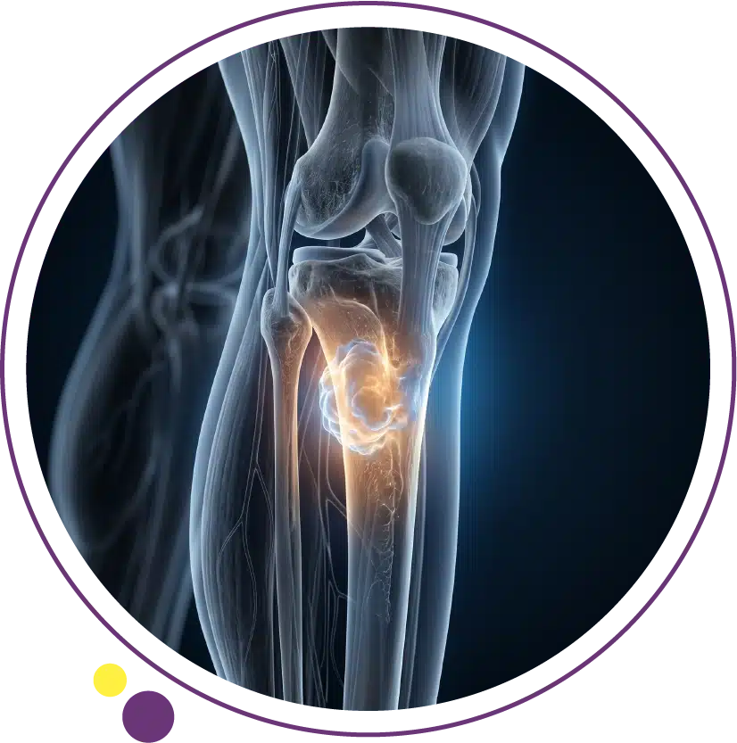
1. Persistent Bone Pain That Worsens Over Time
Bone pain is the most consistent and clinically significant symptom of bone cancer. What distinguishes bone cancer pain from ordinary musculoskeletal pain is its behavior: it is present at rest, it is present at night often waking the person from sleep, it is not reliably relieved by painkillers, and it worsens progressively over weeks and months rather than improving. Ordinary sports injuries and overuse syndromes tend to improve with rest, anti-inflammatory medication, and time. Bone cancer pain does not.
In the earliest stages, the pain may be intermittent and mild, which is exactly why it is so often dismissed or self-treated. The transition from occasional dull aching to constant, severe, and debilitating pain can happen over months. Any bone pain that has been present consistently for more than three to four weeks without a clear mechanical cause, and especially any pain that is worst at night, should prompt imaging. An X-ray is typically the first study requested and can reveal changes in bone density, cortical destruction, or abnormal bony lesions that point toward a more serious underlying process.
2. Swelling and Tenderness Near a Bone
Localised swelling around a bone or joint, particularly in the limbs, is a significant symptom when it appears without a history of injury. Bone tumors can cause surrounding soft tissue to become inflamed and expanded as the tumor grows. The overlying skin may feel warm, and pressing on the area may produce tenderness. In some patients, the swelling is large enough to be visible as a lump beneath the skin. This is particularly common in osteosarcoma, which frequently arises near the knee in the distal femur or proximal tibia.
Swelling near a bone that has appeared without trauma and is associated with pain in the same area should be evaluated clinically and with imaging without delay. An MRI scan provides detailed information about the extent of any soft tissue involvement and the relationship of the tumor to surrounding nerves and vessels, which is critical information for both diagnosis and surgical planning when needed.
3. Weakened Bones and Pathological Fractures
A pathological fracture is a fracture that occurs through bone that has been weakened by disease rather than through normal-strength bone subjected to significant trauma. When a bone tumor destroys normal bone structure, the affected segment becomes mechanically vulnerable. A fracture can occur from a minor fall, a sudden twisting movement, or even during everyday activities that would not ordinarily cause a break in healthy bone.
A pathological fracture is often the first clinical event that leads to a bone cancer diagnosis. It is a serious presentation because it typically indicates that the tumor has been growing for some time before the fracture occurred. Any fracture in a young person without significant trauma, or a fracture in an area that had been painful for weeks before breaking, should trigger investigation for an underlying bone lesion. Plain X-ray of the fracture site will usually reveal the underlying lesion, and further imaging with MRI or CT is then arranged to characterise it fully.
4. Unexplained Fatigue and Weight Loss
Fatigue and unintentional weight loss are non-specific symptoms that accompany many serious illnesses, including bone cancer. They reflect the systemic metabolic demands of a growing tumor on the body. Bone cancer, like other malignancies, can alter normal metabolic processes, suppress appetite, and trigger systemic inflammatory responses that lead to muscle wasting and loss of body weight. These systemic symptoms tend to appear more prominently in aggressive or advanced bone cancers rather than in very early disease.
Unexplained fatigue or weight loss exceeding five to ten percent of body weight over six months, particularly when combined with localised bone pain or swelling, raises the clinical suspicion for malignancy significantly. The imaging services at Images cover the full range of studies needed to evaluate these presentations comprehensively, from initial skeletal X-ray through to staging CT and MRI when a bone lesion is identified.
5. Fever and Night Sweats
Fever and night sweats are more characteristic of certain bone cancer types than others, particularly Ewing sarcoma, which is one of the more common bone cancers in children and young adults. Ewing sarcoma can closely mimic bone infection (osteomyelitis) in its presentation, with fever, localised bone pain, swelling, and an elevated white cell count all appearing together. This clinical overlap makes imaging and biopsy essential for distinguishing between the two conditions.
Night sweats in the context of persistent bone pain and fatigue are a combination that warrants prompt clinical evaluation and imaging. The systemic inflammatory response generated by aggressive bone tumors contributes to both fever and night sweating in some patients. These symptoms alongside bone pain should never be attributed entirely to infection without imaging confirmation of the underlying cause. A CT scan or MRI of the affected area provides far more information than plain X-ray alone in these presentations.
6. Numbness, Tingling, or Limb Weakness
When a bone tumor grows near or presses on a nerve, it can cause neurological symptoms including numbness, tingling, or weakness in the limb served by that nerve. Spinal bone tumors, whether primary or secondary, can compress the spinal cord or nerve roots directly, producing pain that radiates down the leg or arm, sensory disturbance, and in severe cases, significant limb weakness or even bowel and bladder dysfunction. These neurological symptoms represent a more advanced local presentation and typically indicate that urgent imaging and clinical assessment are needed.
Neurological symptoms in the context of bone pain should never be attributed to a benign cause without imaging confirmation. MRI of the spine is the most sensitive tool for evaluating potential spinal cord or nerve root compression and is the study of choice when neurological involvement is suspected alongside a suspected bone lesion. The 3 Tesla MRI service at Images provides the resolution needed to assess these complex presentations accurately.
7. Limited Range of Motion in a Joint
When a bone tumor develops near a joint, it can progressively restrict the normal range of motion in that joint. This occurs as the tumor expands and involves the periarticular structures, causing pain with movement, joint stiffness, and eventually significant limitation in the ability to use the joint normally. The knee, shoulder, and hip are among the most commonly affected joints in primary bone cancer, and loss of full movement in these joints without a clear arthritic or traumatic explanation should prompt investigation.
Joint restriction that develops progressively in a younger patient, particularly in the absence of a prior arthritis diagnosis, is a clinically important finding. Imaging of the joint and adjacent bone typically begins with plain X-ray, and if any bony abnormality is identified, MRI is arranged for detailed characterisation. Our previously published article on diagnostic X-ray explains the role of plain radiography in musculoskeletal evaluation and when follow-up cross-sectional imaging is warranted.
8. A Visible or Palpable Lump Over a Bone
A palpable hard lump directly over a bone that was not previously present is always a significant clinical finding. In some patients, this is the first symptom they notice and the trigger for seeking medical advice. Bone tumors growing outward from the cortex of a bone can produce a hard, fixed mass that can be felt through the skin. The mass may or may not be painful depending on the type of tumor and whether it is compressing surrounding soft tissues or nerves.
Not all bony lumps are malignant. Osteochondromas, which are benign bony outgrowths, are far more common and are typically painless and stable over time. However, any new bony lump that grows, becomes painful, or appears in a young person should be imaged and assessed. The full range of imaging services at Images, including X-ray, MRI, and CT, supports the complete evaluation of any suspected bone lesion from initial detection through to detailed characterisation.
How Symptoms Differ by Bone Cancer Type
Different types of primary bone cancer tend to produce slightly different symptom patterns, partly because of where they arise and the age groups they most commonly affect. Osteosarcoma, the most common primary bone cancer, typically presents in adolescents and young adults with pain and swelling around the knee or shoulder, often initially mistaken for growing pains or sports injury. The pain is frequently worst at night and may precede visible swelling by weeks.
Ewing sarcoma, which is the second most common bone cancer in young people, tends to produce more prominent systemic symptoms including fever, weight loss, and fatigue alongside local bone pain. It can affect any bone but frequently involves the pelvis, femur, and tibia. Chondrosarcoma, which is more common in middle-aged and older adults, often grows more slowly and may produce a longer period of gradually worsening pain before diagnosis. Understanding these different patterns is helpful when the CT scan and MRI findings are being interpreted in their clinical context. For a full breakdown of each bone cancer type, our dedicated article on types of bone cancer covers the key differences in detail.
How Symptoms Vary by Location in the Body
The location of the tumor within the skeleton significantly influences the specific symptoms a patient experiences. Tumors near the knee, which is the most common site for osteosarcoma, produce pain and swelling around the distal femur or proximal tibia, often leading to a limp and difficulty with activities that load the knee. Pelvic tumors may produce deep, diffuse hip and groin pain that is difficult to localize, sometimes causing delays in referral for imaging. Rib cage tumors may present with chest wall pain and difficulty breathing if they grow large enough to impinge on the pleura.
Spinal bone tumors carry the additional risk of cord or nerve root compression, as described above, and may present with back pain combined with neurological symptoms. Skull base tumors, though rare, may present with headache, facial pain, or cranial nerve dysfunction depending on their location and size. For any bone pain that is persistent, progressive, and unexplained by a clear mechanical cause, the appropriate first step is imaging of the affected area. The MRI service at Images provides the detailed soft tissue and bone marrow evaluation needed to characterise lesions in virtually any skeletal location.
When Symptoms Demand Urgent Attention
Certain symptom combinations require urgent medical evaluation rather than a routine appointment. These include sudden severe back pain with any lower limb weakness, bowel or bladder dysfunction, or sensory loss below a specific level, which may indicate acute spinal cord compression requiring emergency imaging and treatment. A fracture through a previously painful bone, or rapidly increasing swelling and pain in a limb in a young person, also warrants same-day assessment.
For less acute but still concerning presentations, persistent unexplained bone pain lasting more than three to four weeks, particularly pain that is worst at night and not improving, should lead to a medical consultation and imaging referral within days rather than weeks. The sooner a bone lesion is identified and characterised, the sooner an appropriate biopsy and treatment plan can be initiated. Patients in Kuwait can arrange X-ray, MRI, and CT scan at any of the Images branches without long waiting times.
What Imaging Reveals That Symptoms Cannot
Symptoms point toward the need for investigation, but imaging provides the objective evidence that turns clinical suspicion into diagnostic certainty. Plain X-ray is the first study to confirm whether a bony lesion is present and to assess its basic characteristics such as location, size, cortical involvement, and any associated abnormal calcification or bone destruction. However, X-ray has limitations in characterising the full extent of a lesion, assessing soft tissue involvement, or detecting lesions in complex anatomical areas such as the pelvis or spine.
MRI provides unparalleled detail for evaluating bone marrow involvement, soft tissue extension, and the relationship of the tumor to surrounding neurovascular structures. It is the modality that most directly determines surgical planning in bone cancer cases. CT scanning contributes to staging by evaluating the lungs and other organs for metastatic spread, and provides detailed three-dimensional bone anatomy for complex cases. Our article on CT scan uses provides broader context on what CT contributes to cancer evaluation beyond what plain X-ray can show. At Images, all three modalities are available across the Jabriya, Hawally, and Salmiya branches to support a complete bone cancer diagnostic workup in Kuwait.
Frequently Asked Questions
Is bone pain at night always a sign of bone cancer?
Not always, but night bone pain is a clinically significant symptom that deserves investigation, particularly when it is persistent and not explained by a recent injury or known condition. Several serious bone conditions including infection, inflammatory arthritis, and both primary and secondary bone tumors can cause nocturnal bone pain. If bone pain consistently disrupts your sleep and has been present for more than a few weeks without improving, a medical consultation and imaging are the appropriate next steps.
Can bone cancer be confused with a sports injury?
Yes, and this is one of the main reasons bone cancer diagnosis is delayed, particularly in young people and athletes. Osteosarcoma near the knee and Ewing sarcoma in the lower limb are both frequently attributed to sports injuries in their early stages because the age groups they affect are physically active and soft tissue injuries are common. The key distinction is that sports injuries improve with rest and physiotherapy, while bone cancer pain persists and worsens. Any limb pain in a young person that does not respond to standard treatment over three to four weeks should prompt X-ray imaging to exclude a bony cause.
Are bone cancer symptoms the same in children and adults?
The core symptoms are similar across age groups, but the types of bone cancer that are most common differ significantly by age. Osteosarcoma and Ewing sarcoma are predominantly seen in children and young adults, while chondrosarcoma is more typical in middle age and beyond. In children, bone cancer may present more prominently with limping, unexplained refusal to use a limb, and pain that is dismissed as growing pains. Any persistent bone pain in a child that disrupts sleep or limits activity warrants imaging.
Can bone cancer spread silently without symptoms?
In early stages, particularly in slower-growing types like chondrosarcoma, the tumor can be present for months before producing noticeable symptoms. This is why imaging that is prompted by any persistent unexplained pain is more likely to catch the disease at a treatable stage than waiting for symptoms to become severe. When bone cancer is found incidentally on imaging performed for another reason, it is often at an earlier and more manageable stage.
What is the first imaging test done when bone cancer is suspected?
Plain X-ray of the affected bone is almost always the first imaging study. It can identify lytic or sclerotic lesions, periosteal reaction, cortical disruption, and abnormal calcification within the lesion. While X-ray is not definitive on its own, it provides the initial evidence that triggers further investigation with MRI or CT. In patients with strongly suspicious clinical and X-ray findings, MRI is usually arranged within days to characterise the lesion in detail before biopsy planning.
Is advanced bone cancer imaging available in Kuwait?
Yes. Images Diagnostic Center provides X-ray, 3 Tesla MRI, and CT scanning across three branches in Kuwait. These modalities together cover the full diagnostic evaluation required for bone cancer investigation, from initial lesion detection through to staging and pre-surgical planning. You can contact the team to arrange imaging at the branch most convenient for you.
Acting on Bone Cancer Symptoms Without Delay
The symptoms of bone cancer are not always dramatic in their early stages, but they are persistent, progressive, and ultimately distinctive when observed over time. Night pain, swelling near a bone without trauma, a fracture from a minor mechanism, and the combination of bone pain with systemic symptoms like fatigue and weight loss are all patterns that deserve serious clinical attention rather than repeated reassurance. Early imaging changes outcomes.
Images Diagnostic Center offers X-ray, MRI, and CT scan services across three accessible branches in Kuwait for patients requiring bone evaluation:
To arrange imaging for a bone concern or to speak with the team about which scan is most appropriate, contact Images directly.