
Lung cancer kills more people globally than any other cancer, and a large part of the reason is timing. Most cases are diagnosed at an advanced stage, when the disease has already spread beyond the lung and curative treatment options are limited. The tragedy is that the lungs themselves do not have pain receptors in their tissue, meaning a tumor can grow for years without producing any sensation that would prompt a person to seek medical attention.
Recognizing the warning signs of lung cancer, understanding why they appear when they do, and knowing when to act on them is one of the most important things a patient or family member can do for their own health. In this article, we will discuss the most important lung cancer symptoms, why they are frequently missed or misattributed, which symptoms should prompt immediate medical attention, how symptoms differ between lung cancer types and stages, and why early imaging is such a critical part of the clinical response when lung cancer is suspected.
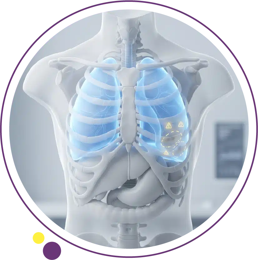
Why Lung Cancer Symptoms Appear Late
The biology of lung cancer creates a fundamental problem for early detection: small tumors in the peripheral lung tissue cause no symptoms at all. The lung parenchyma has no pain fibers, and a tumor can grow to a meaningful size before it presses on adjacent structures, obstructs an airway, or causes any functional change that a patient would notice. By the time symptoms develop, the disease may already be locally advanced or metastatic.
Research consistently shows that the majority of lung cancer patients are diagnosed at stage III or IV, when curative options are significantly more limited than at stage I or II. This is not because patients are ignoring obvious warning signs. It is because the earliest disease produces no signs at all, and even the symptoms of more locally advanced disease are easily attributed to common, less serious conditions like a respiratory infection, acid reflux, or the expected consequences of smoking. Understanding the full picture of what lung cancer looks like at different stages is an important part of our lung cancer overview, which covers the disease from causes through to treatment in comprehensive detail.
The 10 Lung Cancer Symptoms You Should Not Ignore
1. A Persistent Cough That Does Not Go Away
A cough that has lasted for three weeks or more without a clear explanation is one of the most significant and most commonly reported early symptoms of lung cancer. It may be dry or produce mucus, and it often does not feel dramatically different from a cough associated with a respiratory infection, which is exactly why it is so frequently dismissed. The critical distinction is persistence: a cough caused by infection or irritation should resolve or significantly improve within two to three weeks. A cough that does not resolve, that worsens, or that changes in character over several weeks in a patient with risk factors for lung cancer should prompt clinical evaluation and imaging.
Persistent cough in a smoker is particularly easy to explain away as a smoker’s cough, and this normalization delays investigation for many patients. A cough that has changed in quality or frequency, even in a background of chronic smoker’s cough, is not something to dismiss. The CT scan of the chest is the imaging study that provides the detail needed to identify or exclude a lung mass, evaluate the central airways, and assess lymph nodes in patients where a persistent cough has not been explained by other causes.
2. Coughing Up Blood
Hemoptysis, the medical term for coughing up blood, is one of the most alarming symptoms associated with lung cancer and one that should never be normalized or delayed in investigation. Even a small amount of blood in the sputum, sometimes appearing as streaks or a pinkish discoloration, is a serious symptom that requires prompt evaluation. It does not always indicate cancer; other causes including infection, bronchitis, and pulmonary embolism can also cause hemoptysis. But when it occurs in a patient with relevant risk factors, particularly smoking history, it demands immediate clinical attention and imaging.
Central airway tumors, particularly squamous cell carcinoma, are most commonly associated with hemoptysis because they arise near the bronchial blood vessels and can erode into them as they grow. Any episode of coughing up blood warrants chest imaging without delay, typically beginning with a chest X-ray and followed by a CT scan for further characterization if an abnormality is found or if clinical suspicion remains despite a normal X-ray finding. A normal chest X-ray does not exclude a lung tumor, and CT is significantly more sensitive for detecting central and peripheral lesions alike. Our article on diagnostic X-ray explains the relationship between X-ray and CT in chest evaluation and helps patients understand why escalation to CT is sometimes necessary.
3. Unexplained Shortness of Breath
Dyspnea, or breathlessness, that develops without a clear explanation such as a change in fitness level, anemia, or heart failure is a symptom worth investigating carefully in anyone with lung cancer risk factors. A tumor obstructing a bronchus can cause collapse of the part of the lung beyond the obstruction, reducing effective breathing capacity. Fluid accumulating between the lung and the chest wall, called a pleural effusion, is another mechanism by which lung cancer causes breathlessness as the fluid compresses the lung and reduces its ability to expand normally.
Breathlessness that develops gradually and worsens progressively over weeks to months without any other clear cause in a person over 40 with a smoking history should not be attributed to deconditioning or age without imaging investigation. CT of the chest provides detailed evaluation of the lung parenchyma, airways, pleural spaces, and mediastinum in a single study, and is far more sensitive than chest X-ray for identifying the causes of unexplained breathlessness when lung pathology is suspected. The full range of imaging services at Images supports the investigation pathway that begins with symptom assessment and progresses through imaging and specialist review.
4. Chest Pain
Chest pain associated with lung cancer typically reflects involvement of the pleura, the chest wall, the ribs, or the structures of the mediastinum. Unlike the visceral pain of cardiac origin, lung cancer chest pain is often described as a dull, aching, or heaviness in the chest that is present at rest and may worsen with deep breathing or coughing. It can be located in the front or back of the chest and may be difficult for the patient to localize precisely.
A Pancoast tumor, which arises in the apex of the lung and invades the chest wall, brachial plexus, and surrounding structures, can cause a distinctive pain syndrome involving the shoulder, arm, and hand on the affected side, sometimes mimicking orthopedic or neurological conditions for an extended period before the correct diagnosis is established. Any persistent chest pain in a patient with lung cancer risk factors deserves imaging evaluation, and CT of the chest is the appropriate first imaging step for thoracic pain assessment when a pulmonary cause is being considered.
5. Hoarseness
A new or worsening hoarse voice that has persisted for more than two to three weeks without an obvious cause such as a throat infection is a symptom that frequently prompts ENT referral, but it should also prompt consideration of a thoracic cause, particularly in smokers. Lung cancer in the mediastinum can invade or compress the left recurrent laryngeal nerve, which loops down into the chest before returning upward to supply the left vocal cord. When this nerve is damaged, the left vocal cord becomes paralyzed, producing a characteristic hoarse or breathy voice quality.
This symptom reflects a tumor that has already grown beyond the lung into the mediastinum, meaning it represents at least locally advanced disease. The finding of vocal cord paralysis on laryngoscopy should always trigger chest imaging in anyone without another clear explanation. CT of the chest and mediastinum will typically show the causative lesion in these cases. For patients in Kuwait with unexplained hoarseness, timely imaging at Images Diagnostic Center can be arranged promptly to support the clinical assessment of this symptom.
6. Unintentional Weight Loss and Loss of Appetite
Losing significant body weight without intentionally dieting, typically more than five percent of body weight over six to twelve months, is a symptom that should always be investigated regardless of the cause. In the context of lung cancer, weight loss reflects the metabolic demands of a growing tumor combined with cancer-related anorexia and the inflammatory effects of the disease on the body’s nutritional regulation systems. It often accompanies other symptoms and is rarely the sole presenting feature, but it is a meaningful clinical signal that warrants a thorough investigation including chest imaging.
Significant unexplained weight loss in a smoker with any respiratory symptoms is a combination that should prompt early imaging without waiting to see whether weight stabilizes. The sooner a cause is identified, the sooner appropriate investigation and management can be arranged. The CT scan at Images provides the comprehensive chest and abdominal imaging needed to evaluate the possible causes of unexplained weight loss in patients where an oncological process is being considered by the clinical team.
7. Fatigue
Persistent unexplained fatigue is one of the most common and least specific symptoms associated with cancer in general, including lung cancer. It differs from ordinary tiredness in that it is not proportionate to activity levels, does not improve meaningfully with rest, and may be accompanied by a general sense of ill health. Cancer-related fatigue has multiple mechanisms including the metabolic demands of tumor growth, anemia from bone marrow suppression or chronic inflammation, and the systemic effects of inflammatory cytokines released by the tumor or the immune system’s response to it.
While fatigue alone is too non-specific to be a useful trigger for lung cancer investigation, fatigue that occurs alongside respiratory symptoms, weight loss, or cough in a patient with risk factors creates a clinical picture worth taking seriously. If you are concerned about persistent fatigue and its possible causes, speaking with a doctor who can assess the full symptom picture and determine whether imaging is warranted is the appropriate first step. Reviewing our comprehensive article on lung cancer alongside your symptom assessment can help you understand the broader clinical context of what you are experiencing.
8. Recurrent Respiratory Infections
A lung tumor that partially or fully obstructs a bronchus can cause secretions to accumulate behind the obstruction, creating conditions that favor recurrent bacterial infection. Patients may experience repeated episodes of pneumonia or bronchitis that respond to antibiotics but then recur in the same location of the lung. This pattern of recurrent pneumonia in the same lobe or segment is a recognized red flag that should prompt imaging to assess for an obstructing lesion.
Post-obstructive pneumonia is a well-described presentation of central lung cancer, and the infection can mask the underlying tumor on imaging if CT is not performed with appropriate attention to the central airways and the regions beyond the obstructed segment. A chest X-ray may show consolidation that appears infective, but CT provides the cross-sectional detail that reveals whether there is an obstructing mass at the root of the problem. Any patient with two or more episodes of pneumonia in the same lung region within a twelve-month period warrants CT imaging to exclude an underlying structural cause. The CT service at Images is available across all three Kuwait branches for patients requiring this type of thorough chest assessment.
9. Bone Pain
When lung cancer spreads to the skeleton, bone metastases cause pain that is characteristically worse at night, present at rest, and not explained by activity or injury. The spine, ribs, pelvis, and long bones of the limbs are common sites of skeletal metastases from lung cancer. A patient who develops persistent back pain, hip pain, or rib pain without a history of trauma and who has lung cancer risk factors should have this symptom evaluated carefully and not attributed to musculoskeletal cause without appropriate imaging investigation.
When bone metastases are suspected, the diagnostic workup typically includes CT of the chest and abdomen to assess the primary tumor alongside nuclear medicine bone scan or dedicated MRI of symptomatic regions. MRI provides superior detail for assessing bone marrow involvement and identifying cord compression where spinal metastases are present, which can be a clinical emergency requiring urgent treatment. Patients in Kuwait who experience unexplained bone pain alongside any respiratory symptoms should seek prompt medical assessment, and imaging services at Images can support the diagnostic workup their clinician requests.
10. Neurological Symptoms
Lung cancer is one of the most common sources of brain metastases across all cancer types, and neurological symptoms including persistent headache, seizures, visual changes, speech difficulty, weakness on one side of the body, or problems with balance and coordination may be the first indication that lung cancer has spread to the brain. In some patients, neurological symptoms precede the identification of the primary lung tumor entirely, meaning a brain scan prompts chest imaging that leads to the lung cancer diagnosis.
Any new neurological symptom that cannot be explained by a prior neurological condition should prompt brain imaging promptly. MRI of the brain is the most sensitive imaging tool for detecting brain metastases and is preferred over CT for this purpose because of its superior soft tissue contrast and ability to detect smaller lesions. At Images, brain MRI is available using 3 Tesla technology, providing the high-resolution imaging that neurologists and oncologists rely on when assessing intracranial disease in patients with known or suspected lung cancer. For patients who experience anxiety in enclosed scanners, the Open MRI option at Images offers a more comfortable alternative while maintaining the diagnostic quality needed for brain assessment.
Symptoms That Differ Between Lung Cancer Types
The location of the primary tumor within the lung, and whether it arises centrally near the bronchi or peripherally in the lung tissue, significantly influences the symptom pattern a patient experiences. Central tumors, which include most squamous cell carcinomas and small cell lung cancers, tend to cause cough, hemoptysis, and symptoms related to airway obstruction earlier in their course because they arise close to the major breathing tubes. Peripheral tumors, which include most adenocarcinomas, often remain silent for longer because they develop in areas of the lung away from the central airways.
Small cell lung cancer, because of its very rapid growth rate and tendency to spread early, is more likely to present with systemic symptoms such as weight loss and fatigue alongside neurological symptoms from brain metastases or paraneoplastic syndromes, in which the tumor produces hormones or triggers immune responses that affect other organ systems. Paraneoplastic syndromes associated with small cell lung cancer include hyponatremia from inappropriate antidiuretic hormone secretion, and various neurological syndromes. For a detailed explanation of how different lung cancer subtypes behave and how this affects their presentation and management, our article on types of lung cancer provides a thorough breakdown of each major category.
When Symptoms Should Lead to Immediate Action
Some symptom combinations warrant urgent medical attention rather than a routine appointment. These include coughing up a significant amount of blood, a sudden severe neurological symptom such as weakness, speech loss, or seizure, severe and rapidly worsening breathlessness, and acute back pain with any lower limb weakness or bladder disturbance, which may indicate spinal cord compression from a vertebral metastasis. These situations require emergency evaluation and should not be managed by waiting for a scheduled outpatient appointment.
For non-emergency symptoms that are persistent or concerning, the appropriate step is to see a doctor and request imaging. A chest X-ray is typically the first study arranged, but CT provides substantially more diagnostic information and should be requested when clinical suspicion is present, when X-ray findings are inconclusive, or when the initial X-ray is normal but symptoms persist. The imaging services at Images in Kuwait provide CT and MRI options that support the full diagnostic spectrum from initial evaluation through to staging and treatment monitoring. For patients who have been referred for imaging and want to understand what to expect from the CT process, our published guide on types of CT scan provides useful preparation context.
The Importance of Acting on Symptoms Early
Every week of delay between symptom onset and diagnosis represents lost opportunity in lung cancer. The disease that is surgically curable at stage I may be inoperable by the time it reaches stage III, and the progression can occur over months. Patients who act promptly when symptoms appear consistently have more treatment options available, and those options include curative intent therapies that are not available for advanced disease.
This is not about creating unnecessary anxiety or prompting every person with a cough to seek emergency care. It is about understanding which symptom patterns in which patients carry enough clinical significance to warrant investigation, and then acting on that understanding rather than waiting to see if things improve on their own. Patients in Kuwait who are concerned about respiratory symptoms or who have been advised by their doctor to have chest imaging can arrange a CT scan at Images without a long wait, supporting a diagnostic process that is as fast and efficient as the clinical situation demands. The Images health blog provides additional resources on cancer, imaging, and patient guidance for readers who want to stay informed about their health.
Frequently Asked Questions
How early do lung cancer symptoms appear?
In many cases, lung cancer produces no symptoms at all until it has grown to a significant size or has spread to adjacent structures or distant organs. Early-stage peripheral lung cancer is typically asymptomatic. Symptoms become more likely as the tumor grows centrally, obstructs airways, invades the pleura or chest wall, or spreads to lymph nodes and other organs. This is why many patients are diagnosed at an advanced stage and why imaging-based screening in high-risk groups is such an important public health intervention.
Can lung cancer symptoms be mistaken for a chest infection?
Yes, frequently. Cough, fever, breathlessness, and chest discomfort are shared features of lung cancer and respiratory infection. The key distinction is persistence and pattern: infection-related symptoms should improve with appropriate treatment within two to three weeks. Symptoms that do not resolve, that recur repeatedly in the same area, or that are accompanied by significant weight loss or hemoptysis in a patient with risk factors should be investigated with imaging rather than simply treated with repeated courses of antibiotics.
Should I see a doctor if I have a cough for three weeks even without blood?
Yes, particularly if you have lung cancer risk factors such as a history of smoking, significant occupational exposures, or a family history of lung cancer. A persistent cough lasting three weeks or more without a clear and resolving cause warrants clinical evaluation including at minimum a chest examination and likely imaging. Most persistent coughs are caused by non-malignant conditions, but the ones that are not should be identified as early as possible. Your doctor can assess whether a chest X-ray or CT scan is warranted based on your full clinical picture.
Are lung cancer symptoms different in non-smokers?
Non-smoker lung cancer, which is most commonly of the adenocarcinoma subtype, tends to arise in the peripheral lung and may present with minimal or non-specific respiratory symptoms until it has grown substantially. Breathlessness, cough, and chest discomfort can all occur, but because non-smokers may not be considered high-risk for lung cancer, these symptoms may not immediately prompt the same level of investigation. Any persistent unexplained respiratory symptom in a non-smoker should be evaluated if it does not resolve, regardless of the absence of smoking history.
What is the first imaging test done when lung cancer is suspected?
A chest X-ray is usually the first imaging study arranged, providing an overview of the lungs, mediastinum, and chest wall. However, a normal chest X-ray does not exclude lung cancer, particularly for small peripheral tumors. When clinical suspicion remains after a normal X-ray, or when an abnormality is found that needs characterization, a CT scan of the chest is the essential next step. CT is significantly more sensitive and provides the three-dimensional detail needed to characterize any suspected lung lesion properly.
Can imaging alone confirm a lung cancer diagnosis?
No. Imaging can identify a suspicious lesion and characterize its features in ways that help assess the probability of malignancy, but a confirmed cancer diagnosis always requires tissue sampling and pathological examination. CT-guided biopsy, bronchoscopy, or surgical sampling provides the tissue needed for pathological and molecular analysis. The imaging findings guide where and how the biopsy should be performed, and they remain central to staging and monitoring after the tissue diagnosis is established. The Images team can discuss your imaging needs and coordinate efficiently with your referring clinical team.
Recognizing Symptoms Is Only the First Step
The most important message about lung cancer symptoms is this: do not wait. The symptoms discussed in this article are not dramatic or unmistakable in most cases. They are easy to rationalize, easy to postpone, and easy to attribute to something less serious. But for patients whose persistent cough, unexplained breathlessness, or weight loss turns out to reflect an early-stage lung tumor, the decision to seek evaluation promptly rather than waiting can be genuinely life-altering.
If you are concerned about a respiratory symptom that has not resolved, or if you have been advised to arrange chest imaging by your physician, Images Diagnostic Center offers CT scanning and chest imaging across three branches in Kuwait. The imaging team works efficiently with referring physicians to ensure results are available promptly and that the clinical team has the information needed to act without delay.
To arrange your CT scan or speak with the imaging team about your chest health concerns, contact Images directly.