
Lung cancer is the leading cause of cancer-related death worldwide, claiming more lives each year than breast, colon, and prostate cancer combined. Yet it is also a disease that is increasingly understood, increasingly treatable, and in more cases than ever before, detectable at a stage where meaningful intervention is possible.
For patients, families, and anyone who wants to understand what lung cancer actually involves, having accurate and complete information matters enormously. The decisions made in the weeks following a diagnosis, or in the period before a diagnosis when symptoms first appear, can significantly affect outcomes. In this article, we will discuss what lung cancer is, how it develops, the main types and their differences, the most common risk factors and causes, how symptoms present and why they are often missed, how lung cancer is diagnosed and staged, what treatment options are available, and why early imaging plays such a critical role in improving outcomes.
What Is Lung Cancer?
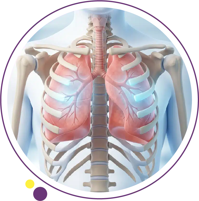
Lung cancer begins when cells in the lungs mutate and begin to grow in an uncontrolled way, forming a tumor that can invade surrounding tissue and spread to other parts of the body through the blood or lymphatic system. The lungs are two large, spongy organs in the chest responsible for exchanging oxygen and carbon dioxide with every breath. Because they are so central to basic physiology, tumors growing within them can disrupt breathing, cause bleeding into the airways, and obstruct the structures around them as they enlarge.
Lung cancer is broadly divided into two main categories based on how the cancer cells appear under a microscope: non-small cell lung cancer and small cell lung cancer. These two categories behave very differently, spread at different rates, and are treated with different approaches. Understanding which type a patient has is one of the first and most important steps in the diagnostic process. The CT scan is the primary imaging tool used to detect, characterize, and stage lung cancer, and it plays a role at virtually every point in the diagnostic and treatment pathway.
Risk Factors and Causes of Lung Cancer
Smoking
Tobacco smoking is the single most important risk factor for lung cancer and is responsible for approximately 85 percent of all cases globally. The risk is directly proportional to how long a person has smoked and how many cigarettes they have smoked per day. Even after stopping, the elevated risk persists for years, though it gradually diminishes over time. Tobacco carcinogens damage the DNA of lung cells over years of repeated exposure, and the accumulated mutations eventually overcome the cell’s normal control mechanisms. Passive exposure to secondhand smoke also increases risk, though less markedly than active smoking.
Former and current smokers who meet specific age and smoking history criteria are now considered candidates for annual low-dose CT lung screening in many international guidelines. This approach has been shown to reduce lung cancer mortality by detecting tumors when they are smaller and more treatable. The imaging services at Images include CT scanning that can be used as part of a clinician-directed lung assessment, and patients with a significant smoking history should discuss the possibility of lung cancer screening with their doctor.
Radon Gas
Radon is a naturally occurring radioactive gas that forms from the decay of uranium in soil and rock. It can accumulate in enclosed spaces, particularly in basements and lower floors of buildings, and is the second leading cause of lung cancer after smoking in many countries. Because it is colorless and odorless, radon cannot be detected without specific testing. Exposure is cumulative, and the combination of radon exposure and smoking creates a compounded risk that is substantially higher than either factor alone. Radon awareness is growing internationally, and testing is increasingly recommended for homes in areas where geological conditions favor higher radon levels.
For patients in Kuwait investigating respiratory symptoms or lung findings discovered incidentally on imaging, understanding the full range of risk factors is an important part of the clinical conversation with a specialist. The CT scan is the most sensitive tool for characterizing lung nodules identified on initial chest imaging, and early follow-up CT is often recommended when a lung abnormality is found on a chest X-ray.
Occupational Exposures and Other Risk Factors
Occupational exposure to substances such as asbestos, arsenic, chromium, nickel, beryllium, and diesel exhaust increases lung cancer risk, particularly in workers who have had prolonged contact over years or decades. Asbestos exposure is particularly significant because it acts synergistically with smoking to multiply risk. Air pollution, including fine particulate matter from vehicle emissions and industrial activity, is recognized as a contributing environmental risk factor for lung cancer, particularly in densely populated urban areas.
A family history of lung cancer confers a modestly elevated risk independent of smoking, suggesting that genetic susceptibility plays a role in some cases. Certain lung diseases such as pulmonary fibrosis and chronic obstructive pulmonary disease also appear to increase lung cancer risk beyond what is explained by smoking history alone. Patients who have multiple risk factors should discuss screening and monitoring with their physician. Understanding the broader landscape of cancer diagnosis pathways helps patients appreciate how symptoms, risk factors, and imaging findings are connected in the clinical evaluation process.
How Lung Cancer Develops and Spreads
Lung cancer does not develop overnight. In most cases, years or even decades of cumulative cellular damage precede the emergence of a clinically detectable tumor. Repeated exposure to carcinogens gradually damages the DNA of bronchial epithelial cells, leading to mutations that accumulate over time until the cell’s regulatory mechanisms are sufficiently disrupted that uncontrolled growth begins.
Once a primary tumor forms, lung cancer can spread locally by invading adjacent structures including the pleura, chest wall, mediastinum, heart, and great vessels. It can spread regionally by travelling through the lymphatic system to hilar and mediastinal lymph nodes. And it can spread distantly through the bloodstream to organs including the brain, liver, adrenal glands, bones, and the opposite lung. The extent of spread at the time of diagnosis is the primary determinant of stage, which in turn is the primary driver of treatment options and outcomes. The MRI scan is particularly important in assessing brain metastases in lung cancer patients, as MRI provides superior sensitivity for intracranial lesions compared to CT, and brain involvement significantly influences staging and treatment planning.
Types of Lung Cancer
Non-Small Cell Lung Cancer
Non-small cell lung cancer accounts for approximately 85 percent of all lung cancer diagnoses and encompasses several distinct subtypes including adenocarcinoma, squamous cell carcinoma, and large cell carcinoma. Adenocarcinoma is the most common subtype overall and tends to arise in the peripheral lung tissue rather than the central airways. It is the most frequent type seen in non-smokers and is characterized by specific molecular mutations that make it particularly amenable to targeted therapy.
Squamous cell carcinoma typically arises in the central airways near the bronchi and is closely associated with smoking history. Large cell carcinoma is a less common subtype characterized by rapidly growing tumors without the distinguishing features of adenocarcinoma or squamous cell carcinoma under the microscope. Non-small cell lung cancer generally grows and spreads more slowly than small cell lung cancer, and surgical resection remains a curative option for patients diagnosed at an early localized stage. The CT scan of the chest is the essential imaging study for characterizing the primary tumor, its relationship to surrounding structures, and the regional lymph nodes before any treatment decision is made. For a more detailed breakdown of how lung cancer subtypes differ in behavior and management, our dedicated article on types of lung cancer covers each subtype in full.
Small Cell Lung Cancer
Small cell lung cancer accounts for approximately 15 percent of lung cancers and is almost exclusively associated with tobacco smoking. It is a highly aggressive cancer that tends to grow rapidly and spread to distant sites early in its course, meaning many patients have advanced disease at the time of diagnosis. Small cell lung cancer is classified as either limited stage, confined to one side of the chest, or extensive stage, spread beyond one hemithorax or to distant organs.
Despite its aggressive nature, small cell lung cancer is initially very sensitive to chemotherapy and radiation, and many patients achieve significant responses to first-line treatment. However, relapse is common, and long-term outcomes for extensive stage disease remain challenging. Immunotherapy has recently been added to first-line chemotherapy regimens for extensive stage small cell lung cancer, improving outcomes modestly but meaningfully. Brain metastases are particularly common in small cell lung cancer, and prophylactic cranial irradiation is used in some patients who achieve a good response to initial treatment. Regular imaging with CT and MRI of the brain forms the backbone of monitoring and response assessment throughout treatment for this aggressive disease.
Symptoms of Lung Cancer
One of the most challenging aspects of lung cancer is that it frequently produces no symptoms in its early stages. The lungs have no pain receptors in their parenchyma, and a small tumor growing in peripheral lung tissue can be present for years without causing any noticeable symptom. By the time symptoms do develop, the disease is often locally advanced or metastatic, which significantly narrows treatment options and affects outcomes.
Common symptoms of lung cancer include a persistent cough that does not resolve over several weeks, coughing up blood even in small amounts, unexplained shortness of breath, chest pain, hoarseness, recurrent respiratory infections, unintentional weight loss, and fatigue. When cancer has spread to the bones, bone pain may be prominent. When it has spread to the brain, headaches, visual changes, or neurological symptoms may develop. None of these symptoms is specific to lung cancer, but their persistence and combination in a person with relevant risk factors should always prompt clinical evaluation including imaging. For a detailed breakdown of the specific warning signs and their clinical significance, our article on lung cancer symptoms provides a thorough guide to help patients recognize which patterns warrant urgent attention.
How Lung Cancer Is Diagnosed
The diagnostic process for lung cancer typically begins when a lung abnormality is identified, either because a patient presents with symptoms that prompt imaging, or because a nodule or mass is found incidentally on a scan performed for another reason. A chest X-ray may be the first study to suggest a lung lesion, but CT scanning of the chest is always required for proper characterization. CT provides cross-sectional imaging of the lung parenchyma, mediastinum, and chest wall with a level of detail that plain X-ray cannot approach, and it is the standard imaging study for evaluating any suspected lung mass or significant lung abnormality. Our article on CT scan uses explains in detail why CT is the tool of choice for chest and lung assessment in this context.
Once a suspicious lesion is identified on CT, tissue sampling is required to confirm the diagnosis, determine the cancer type, and obtain material for molecular profiling. Bronchoscopy is used when tumors are located near the central airways and can be accessed through the breathing tubes. CT-guided needle biopsy is used for peripheral lung lesions that can be accessed percutaneously. In some cases, lymph node sampling via endobronchial ultrasound-guided biopsy is performed to assess mediastinal involvement. The pathological and molecular analysis of the biopsy sample is as important as the imaging, because it determines which treatment options are available to the individual patient.
For patients in Kuwait who require CT imaging of the chest, either for initial assessment of a lung concern or for follow-up of a known finding, the CT scan service at Images is available across all three branches. Timely access to imaging is an important part of avoiding diagnostic delays that can affect treatment options and outcomes.
Staging Lung Cancer
Staging lung cancer defines how far the disease has spread and is the framework that determines which treatment approaches are appropriate. The TNM staging system used for non-small cell lung cancer assesses three dimensions: T for the size and local extent of the primary tumor, N for regional lymph node involvement, and M for the presence of distant metastases. These combine to produce an overall stage from I through IV, with stage I representing localized disease confined to the lung and stage IV representing metastatic disease.
Staging requires comprehensive imaging that extends beyond the chest. CT of the chest and abdomen assesses the primary tumor, lymph nodes, liver, adrenal glands, and other abdominal structures. MRI of the brain is recommended in patients with stage III or IV disease, symptoms suggesting intracranial involvement, or in patients with small cell lung cancer regardless of stage, because brain metastases are common and their presence or absence significantly influences the overall treatment plan. Bone assessment may also be required when bone pain or elevated calcium levels suggest skeletal involvement. Accurate staging is only possible with complete and high-quality imaging, and the full range of imaging services at Images supports this comprehensive assessment for patients in Kuwait.
Treatment Options for Lung Cancer
Surgery
Surgical resection is the primary curative treatment for early-stage non-small cell lung cancer when the tumor is confined to the lung and the patient is fit enough to tolerate the procedure. The most common surgical approach is a lobectomy, removing the lobe of the lung containing the tumor. In patients with limited pulmonary reserve, a more limited resection such as a segmentectomy or wedge resection may be considered. Video-assisted thoracoscopic surgery has reduced the invasiveness of many lung cancer operations compared to traditional open thoracotomy, shortening recovery time and reducing complications in appropriate cases.
Pre-surgical imaging is essential to confirm the tumor’s location, relationship to surrounding structures, lymph node status, and the condition of the opposite lung. CT provides the anatomical roadmap for surgical planning, and MRI or additional imaging may be used when specific structures are of concern. Post-surgical CT imaging is used to confirm the adequacy of resection and to monitor for local recurrence during follow-up. The CT service at Images supports both pre-operative assessment and post-treatment monitoring for patients in Kuwait managing lung cancer.
Radiation Therapy
Radiation therapy uses high-energy beams to destroy cancer cells and is used in lung cancer in several contexts. Stereotactic body radiotherapy delivers highly focused, high-dose radiation to small peripheral lung tumors and is increasingly used as a curative alternative to surgery for patients with early-stage disease who are not surgical candidates. Conventionally fractionated radiation combined with chemotherapy is the standard approach for locally advanced stage III non-small cell lung cancer that cannot be surgically removed. In small cell lung cancer, concurrent chemoradiotherapy is the standard treatment for limited stage disease. Palliative radiation is used to relieve symptoms caused by the primary tumor or metastases, including superior vena cava obstruction, bone pain, and in some cases brain metastases.
Radiation planning relies heavily on imaging, including CT simulation and in some cases MRI, to precisely define the treatment target and spare surrounding normal tissues. For patients receiving radiation for lung cancer in Kuwait, MRI of the brain and CT imaging before and after treatment provide the staging and response data that radiation oncologists and medical oncologists need for coordinated care planning. The Images health blog covers imaging guidance relevant to patients across different cancer types and treatment stages.
Chemotherapy
Chemotherapy remains an important component of lung cancer treatment across multiple settings: as adjuvant therapy after surgery to reduce relapse risk, as part of concurrent treatment with radiation for locally advanced disease, and as systemic therapy for advanced or metastatic disease. Platinum-based doublet chemotherapy has been the backbone of first-line treatment for advanced non-small cell lung cancer for many years and remains relevant even as targeted and immunotherapy options have expanded, particularly for patients whose tumors lack targetable mutations.
For small cell lung cancer, chemotherapy with or without immunotherapy remains the standard systemic treatment. Response to chemotherapy is assessed through clinical evaluation and regular CT imaging, which measures changes in tumor size and the emergence of new lesions across the treatment course. Patients in Kuwait receiving lung cancer treatment can access CT and MRI imaging at Images to support the monitoring their oncologists require throughout and after their treatment program.
Targeted Therapy
Targeted therapies have transformed outcomes for a significant subset of lung cancer patients, particularly those with adenocarcinoma whose tumors carry specific molecular mutations. EGFR mutations are present in approximately 10 to 15 percent of Western lung adenocarcinoma patients and in higher proportions in East Asian populations, and EGFR tyrosine kinase inhibitors produce high response rates in this population. ALK and ROS1 rearrangements are present in smaller proportions of patients and are effectively targeted by specific inhibitors. KRAS G12C mutations, previously considered untargetable, can now be addressed with sotorasib and related agents.
Molecular testing of lung cancer tissue is now a standard part of the diagnostic workup for advanced non-small cell lung cancer because the results directly determine which targeted therapies are applicable. This makes the quality and adequacy of the initial biopsy critically important. Imaging monitoring with CT throughout targeted therapy treatment tracks response, identifies when resistance is developing, and guides decisions about changing therapy. The CT scan is the primary tool for this ongoing assessment.
Immunotherapy
Immune checkpoint inhibitors, particularly agents targeting the PD-1 and PD-L1 pathways, have become an established part of lung cancer treatment for both non-small cell and small cell subtypes. PD-L1 expression on tumor cells is tested at diagnosis to predict the likelihood of response, with high expressers showing particularly favorable responses to pembrolizumab and related agents. Immunotherapy may be used as monotherapy in first-line treatment of PD-L1 high non-small cell lung cancer, or in combination with chemotherapy across a broader range of patients. Atezolizumab and durvalumab have specific approvals in small cell and locally advanced non-small cell lung cancer respectively.
Immunotherapy responses differ from chemotherapy responses in that some patients achieve very durable control that extends well beyond the treatment period. Imaging monitoring during and after immunotherapy requires awareness of specific patterns such as pseudoprogression, where tumors may appear temporarily larger before responding, and immune-related adverse events that can affect the lungs, liver, and other organs. Regular imaging remains essential throughout immunotherapy, and the imaging services at Images in Kuwait are suited to supporting patients receiving these newer treatment approaches.
The Critical Importance of Early Detection
The relationship between lung cancer stage at diagnosis and survival is stark and direct. Five-year survival rates for stage I lung cancer, where the disease is localized to the lung, are substantially higher than for stage IV disease, where distant metastases are present. The challenge is that stage I lung cancer typically produces no symptoms, meaning that without active screening or incidental detection on imaging, most patients are diagnosed only when symptoms prompt clinical evaluation, at which point advanced disease is often already present.
This reality is what makes low-dose CT lung screening such an important intervention for high-risk individuals. Multiple large clinical trials have demonstrated that annual low-dose CT screening in current and former heavy smokers reduces lung cancer mortality by detecting cancers at earlier and more treatable stages. For individuals who qualify based on age and smoking history, discussing CT screening with their physician is a genuinely important health decision. The CT scan service at Images is available to patients in Kuwait for clinician-directed lung assessment, and early referral for imaging when respiratory symptoms arise is equally important for those who do not qualify for formal screening programs. Patients interested in understanding more about the diagnostic processes involved in cancer detection can explore our overview of cancer diagnosis for a broader perspective on how imaging, biopsy, and staging work together.
Frequently Asked Questions
Can lung cancer develop in people who have never smoked?
Yes. Approximately 10 to 15 percent of lung cancer cases globally occur in people who have never smoked. Non-smoker lung cancer is more commonly of the adenocarcinoma subtype and is more frequently associated with specific molecular mutations such as EGFR or ALK that may have different causative pathways including genetic susceptibility, radon exposure, air pollution, and secondhand smoke exposure over many years. Non-smokers with lung cancer symptoms or lung findings on imaging should receive the same thorough evaluation as smokers.
What is the most important scan for diagnosing lung cancer?
CT scanning of the chest is the most important imaging study for evaluating suspected lung cancer. It characterizes the primary tumor, assesses lymph node involvement, and identifies additional lung nodules or local complications in far more detail than a chest X-ray. CT of the abdomen is added for staging purposes, and MRI of the brain is performed in many patients to assess for intracranial metastases that would affect staging and treatment decisions.
Is lung cancer treatable if found early?
Yes, significantly so. Early-stage lung cancer confined to the lung is potentially curable with surgical resection or, in patients who are not surgical candidates, with stereotactic body radiotherapy. The challenge is that early lung cancer is usually asymptomatic, making early detection dependent on screening or incidental imaging findings. When lung cancer is found at stage I or II, the prognosis and treatment options are substantially better than for advanced disease, which underscores the importance of prompt investigation when risk factors or symptoms are present.
How is lung cancer monitored during and after treatment?
CT scanning is the standard monitoring tool throughout lung cancer treatment, used to measure tumor response, detect new lesions, and identify complications or disease progression. MRI is used for brain monitoring in patients at risk of intracranial metastases. After treatment completion, follow-up CT scans are scheduled at regular intervals for several years to detect recurrence early. Patients in Kuwait can arrange their follow-up CT scans at any of the three Images branches, with results communicated promptly to the referring oncologist or specialist.
What should someone do if they notice possible lung cancer symptoms?
Anyone who notices a persistent cough lasting more than three weeks, blood in sputum, unexplained breathlessness, chest pain, or significant unintentional weight loss should see a doctor promptly rather than waiting to see if symptoms resolve on their own. These symptoms do not confirm lung cancer, but they warrant clinical evaluation that typically includes a chest X-ray and, where indicated, a CT scan. Early investigation is far better than delayed diagnosis. Familiarizing yourself with the full range of lung cancer symptoms can help you identify which specific signs deserve prompt medical attention.
Is CT lung screening available in Kuwait?
CT scanning for lung assessment is available at Images Diagnostic Center across three branches in Kuwait. For individuals who are considering lung screening based on smoking history or other risk factors, the most appropriate first step is to discuss this with their physician, who can assess eligibility based on clinical criteria and arrange a referral. The Images team can then coordinate the CT scan efficiently once a referral is in place.
Taking the Next Step
Lung cancer is serious, but it is not without options. Early detection, accurate staging, molecular testing, and access to the right treatment for the right patient at the right stage are the pillars that define outcomes. The role of imaging throughout this process is indispensable, from the first CT scan that characterizes a suspicious finding, through the staging workup, the treatment response assessments, and the long-term follow-up surveillance that keeps recurrence from going undetected.
For patients in Kuwait who need chest or lung imaging, brain assessment, or any other scan related to a lung cancer investigation or treatment program, Images Diagnostic Center provides CT and MRI services across three accessible branches. Timely imaging access reduces the delays that matter most in an oncological context, and the team at Images is equipped to coordinate efficiently with referring physicians and oncology specialists.
To arrange a CT scan, MRI, or any imaging related to lung cancer assessment in Kuwait, contact the Images team and they will help coordinate the right scan for your clinical needs.