
A blood cancer diagnosis changes everything, and one of the first questions most patients and families ask is: what are the treatment options? The answer is more hopeful than it used to be. Advances in oncology over the past two decades have transformed blood cancer treatment, with many patients achieving long-term remission or manageable chronic disease on therapies that were not available a generation ago.
Understanding how blood cancer is treated, what determines which approach is right for a given patient, and what the process of care looks like from diagnosis through to monitoring is an important part of navigating this experience with clarity. In this article, we will discuss the main types of blood cancer and how their treatment differs, the key therapeutic approaches including chemotherapy, targeted therapy, immunotherapy, and stem cell transplantation, how diagnosis and staging guide treatment decisions, the role of imaging in monitoring treatment response, and what patients can expect at each stage of care.
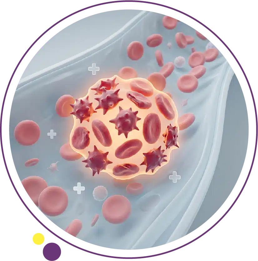
Understanding the Different Types of Blood Cancer
Blood cancer is not a single disease. It is a broad term covering a group of cancers that originate in the blood, bone marrow, or lymphatic system and affect how blood cells are produced, function, and multiply. The three main categories are leukemia, lymphoma, and myeloma, each of which behaves differently, presents with different symptoms, and requires a different treatment approach. For a detailed breakdown of how each type develops and is classified, our article on blood cancer types covers the key distinctions in accessible detail.
Leukemia involves the overproduction of abnormal white blood cells within the bone marrow, which crowds out normal blood cell production. It is divided into acute and chronic forms, and further classified by the type of white cell affected: lymphoid or myeloid. Lymphoma arises in the lymphatic system and is categorized as Hodgkin or non-Hodgkin lymphoma, with non-Hodgkin lymphoma covering a large and diverse group of subtypes. Multiple myeloma is a cancer of plasma cells within the bone marrow and is associated with bone pain, kidney impairment, and abnormal protein production. Each of these categories responds differently to treatment, which is why the type and subtype of blood cancer is the most fundamental factor in determining the appropriate therapeutic plan. Understanding the cancer diagnosis process fully equips patients to engage meaningfully with the treatment planning that follows.
How Blood Cancer Is Diagnosed and Staged Before Treatment
Treatment planning for blood cancer begins with a comprehensive diagnostic workup that goes well beyond a basic blood test. Accurate diagnosis requires bone marrow biopsy in most cases, along with detailed pathological and genetic analysis of the cancer cells. This molecular and cytogenetic profiling is not a formality; it directly determines which treatments are most likely to be effective and which the cancer is most likely to resist.
Staging identifies how widely the cancer has spread and assesses how it is affecting the body’s normal function. For lymphomas, staging uses imaging to assess lymph node involvement and organ spread. CT scanning is routinely used to identify enlarged lymph nodes, assess spleen and liver size, and detect masses in the chest, abdomen, or pelvis. In many lymphoma cases, PET-CT is used alongside CT to detect metabolically active disease. At Images, CT scan is available across all three Kuwait branches to support both initial staging assessments and follow-up monitoring throughout the treatment course.
For myeloma, imaging of the spine, pelvis, and long bones is an important component of staging and disease monitoring. MRI of the spine is particularly valuable in myeloma because it identifies bone marrow infiltration and assesses for cord compression that may require urgent intervention, providing detail that plain X-ray cannot match for marrow-based disease assessment.
Chemotherapy in Blood Cancer Treatment
Chemotherapy remains one of the foundational treatments for many types of blood cancer, particularly acute leukemias and aggressive lymphomas where rapid disease control is the immediate priority. It works by targeting rapidly dividing cells, including cancer cells, and is typically given in cycles with rest periods in between to allow normal cells to recover. In acute leukemia treatment, chemotherapy is given in distinct phases: induction, which aims to achieve remission, consolidation, which aims to deepen and sustain remission, and in some cases maintenance therapy to prevent relapse.
Combination chemotherapy regimens, using multiple drugs together, are standard in most blood cancer treatment protocols because they target cancer cells through different mechanisms simultaneously, reducing the likelihood of treatment resistance developing. The specific drugs, doses, and schedules used depend entirely on the cancer type and subtype, the patient’s fitness and organ function, and whether the treatment is being given with curative or disease-control intent. Side effect management is an important part of chemotherapy care, and patients are closely supported through treatment with antiemetics, growth factors to support blood cell recovery, and infection prevention strategies. For patients and families building their foundational understanding of blood cancer, reviewing the known causes of blood cancer alongside the treatment landscape provides useful context for discussions with the oncology team.
Targeted Therapy: Precision Treatment for Blood Cancer
Targeted therapies represent one of the most significant advances in blood cancer treatment over the past twenty years. Unlike chemotherapy, which affects all rapidly dividing cells, targeted therapies are designed to interfere with specific molecular pathways or genetic mutations that drive cancer cell growth in particular blood cancer subtypes. This precision means they can be highly effective against specific cancers while sparing many normal tissues, often resulting in a more favorable side effect profile compared to conventional chemotherapy.
Tyrosine kinase inhibitors are among the most well-known targeted therapies for blood cancer. Imatinib, the first of this class, transformed the treatment of chronic myeloid leukemia (CML) with the BCR-ABL mutation from a disease requiring stem cell transplant to one manageable with daily oral medication for most patients. Successive generations of tyrosine kinase inhibitors have extended these benefits and addressed resistance patterns that emerged with earlier agents.
Venetoclax targets the BCL-2 protein that many blood cancer cells rely on to evade programmed cell death and is used in chronic lymphocytic leukemia and certain types of acute myeloid leukemia. BTK inhibitors such as ibrutinib are effective in B-cell malignancies including CLL and certain lymphoma subtypes. The expanding landscape of targeted options means that molecular profiling at diagnosis is increasingly central to treatment planning, as the presence or absence of specific mutations directly determines which targeted agents are likely to be effective. Imaging plays a supporting role in monitoring response to these therapies, and the full imaging services at Images are available to support the ongoing assessment that patients undergoing blood cancer treatment require.
Immunotherapy and Blood Cancer
Immunotherapy works by harnessing or enhancing the patient’s own immune system to recognize and destroy cancer cells. Several forms of immunotherapy are now established in blood cancer treatment, each working through different mechanisms.
Monoclonal Antibodies
Monoclonal antibody therapy uses laboratory-engineered proteins that specifically bind to antigens on the surface of cancer cells, marking them for destruction by the immune system or directly blocking growth signals. Rituximab, which targets the CD20 protein on B cells, has become a cornerstone of treatment for most B-cell lymphomas and is combined with chemotherapy in many standard regimens. Obinutuzumab is a next-generation CD20 antibody used in certain lymphoma and CLL protocols. Daratumumab targets CD38 on myeloma cells and is now used across multiple lines of myeloma therapy.
These agents are generally administered by intravenous infusion, often alongside chemotherapy, and have substantially improved outcomes for many blood cancer patients over the past two decades. Monitoring the response to antibody-based therapies involves both clinical assessment and regular imaging to track disease burden. The CT scan service at Images supports this monitoring function for patients managing blood cancer in Kuwait, providing the imaging evidence that oncologists need to assess treatment efficacy and inform the next step in care.
CAR-T Cell Therapy
Chimeric antigen receptor T cell therapy, known as CAR-T, is one of the most significant recent developments in blood cancer treatment. It involves extracting a patient’s own T cells, genetically engineering them to express receptors that target specific cancer cell antigens, and then reinfusing them after expansion in the laboratory. CAR-T cells can achieve dramatic responses in patients with relapsed or refractory B-cell lymphoma and certain leukemias who have failed multiple lines of prior treatment.
CAR-T therapy is currently available at specialized cancer centers and requires careful monitoring for specific toxicities including cytokine release syndrome and neurological effects. It represents a genuine step forward for patients who have exhausted conventional options, and its role in earlier lines of therapy is under active investigation in clinical trials. The complex nature of CAR-T treatment requires ongoing imaging assessment as part of response evaluation, and patients followed in Kuwait for blood cancer management can arrange their CT or MRI imaging needs through Images Diagnostic Center as part of their broader care pathway.
Checkpoint Inhibitors
Checkpoint inhibitors block proteins that cancer cells use to shield themselves from immune attack. Pembrolizumab and nivolumab, which target the PD-1 checkpoint pathway, have shown significant activity in classical Hodgkin lymphoma and are approved for use in relapsed or refractory disease. Their role in other blood cancers is an active area of research. While checkpoint inhibitors are better established in solid tumors, their application in haematological malignancies continues to expand as clinical trial evidence accumulates. Response monitoring with CT imaging is an integral part of managing patients receiving these therapies, assessing whether lymph node masses are shrinking or whether new disease sites are appearing during treatment.
Radiation Therapy in Blood Cancer
Radiation therapy plays a more limited but still meaningful role in blood cancer treatment compared to solid tumors. In Hodgkin lymphoma, radiation to involved lymph node regions following chemotherapy has historically been part of combined modality treatment and remains used in certain presentations. In non-Hodgkin lymphoma, localized radiotherapy can be effective for certain indolent subtypes or as palliative treatment for bulky disease causing symptoms. In multiple myeloma, targeted radiation is used to treat painful bone lesions or to address cord compression where urgent intervention is needed.
The decision to include radiation in a blood cancer treatment plan depends on the specific disease, its extent, the patient’s age and prior treatment, and the goals of care. Radiation planning requires detailed imaging of the treatment area, and the imaging performed before and after radiotherapy is an essential component of treatment response assessment. Patients in Kuwait undergoing blood cancer care who require staging or response assessment imaging can access both CT and MRI services at Images to support their clinical team’s decision-making at each stage of treatment.
Stem Cell Transplantation
Stem cell transplantation remains a central treatment option for many patients with blood cancers that require intensive therapy to achieve or sustain remission. An autologous stem cell transplant uses the patient’s own stem cells collected before high-dose chemotherapy, which is then used to rescue the bone marrow after treatment. This approach is used in myeloma and certain lymphoma cases. An allogeneic transplant uses stem cells from a matched donor, either related or unrelated, and offers not only bone marrow rescue but also an immune graft-versus-leukemia effect in some patients, which can contribute to long-term disease control.
Transplantation carries significant risks including infection, graft-versus-host disease in the allogeneic setting, and organ toxicity from conditioning regimens. Patient selection, timing, and the availability of suitable donors or stored cells are all carefully assessed before transplant is recommended. Pre-transplant evaluation includes comprehensive imaging to confirm disease status before proceeding, and post-transplant monitoring includes regular follow-up imaging to detect relapse or complications. The MRI and CT services at Images support both pre-transplant staging and post-treatment monitoring for patients managed in Kuwait.
The Role of Imaging in Blood Cancer Treatment Monitoring
Imaging is not only relevant at diagnosis and staging. It plays an ongoing and critical role throughout blood cancer treatment and long-term follow-up. CT scanning is the standard tool for assessing lymph node response, tracking disease in the chest and abdomen, and detecting relapse after treatment in lymphoma. MRI provides superior soft tissue detail for myeloma monitoring and is the preferred modality for assessing spinal cord and bone marrow involvement.
Interim imaging studies performed during treatment allow oncologists to assess whether the cancer is responding as expected or whether the treatment approach needs to be adjusted early. End-of-treatment imaging confirms the depth of response achieved and informs decisions about consolidation, maintenance, or surveillance. Follow-up imaging schedules in the years after treatment aim to detect relapse before symptoms develop, enabling prompt intervention when disease recurs. For patients managing blood cancer in Kuwait, having reliable access to high-quality imaging at a local center reduces the practical burden of this ongoing monitoring. The imaging services at Images across Jabriya, Hawally, and Salmiya are accessible precisely for this purpose, supporting the clinical teams and patients who need consistent and timely imaging throughout a prolonged treatment journey.
Supportive Care and Managing Treatment Side Effects
Effective blood cancer treatment involves far more than the primary anti-cancer therapies. Supportive care addresses the side effects and complications that arise during treatment and significantly influences the patient’s ability to complete their planned therapy and maintain quality of life throughout. Blood transfusions and platelet transfusions may be needed to manage cytopenias caused by chemotherapy or bone marrow suppression from the disease itself. Growth factor injections stimulate white blood cell recovery to reduce infection risk during chemotherapy.
Infection prevention and prompt treatment are particularly important in blood cancer patients because immune function is often compromised both by the disease and by the treatments used. Nutritional support, pain management, psychological support, and physiotherapy all form part of the broader care model that specialist haematology teams coordinate. Fatigue, neuropathy, mucositis, and other treatment effects are common and are managed with targeted interventions rather than accepted as inevitable. Patients in Kuwait accessing blood cancer care are supported by multidisciplinary teams, and imaging at Images Diagnostic Center integrates naturally into this care model when staging, monitoring, or complication assessment is required. For patients beginning to understand the broader landscape of blood cancer, reviewing the cancer diagnosis overview is a practical resource for understanding how the diagnostic and treatment phases connect.
Frequently Asked Questions
Is blood cancer curable?
Many types of blood cancer are curable with current treatment, and the prognosis depends significantly on the specific type and subtype, the stage at diagnosis, the molecular characteristics of the cancer, and the patient’s overall health. Hodgkin lymphoma, for example, has a high cure rate with modern treatment. Many cases of acute leukemia in younger patients are cured with intensive chemotherapy, sometimes followed by stem cell transplantation. Others, such as myeloma, are currently considered chronic conditions that can be managed effectively over many years rather than cured outright. Early, accurate diagnosis and prompt treatment planning give the best chance of achieving the deepest possible response.
How is the right blood cancer treatment chosen?
Treatment selection is based on several factors: the specific type and molecular subtype of blood cancer, the extent of disease at diagnosis, the patient’s age and fitness, organ function, prior treatments if this is a relapse, and the goals of care. Molecular and genetic profiling of the cancer cells at diagnosis is now central to this decision in most blood cancer types, as it identifies which targeted therapies are likely to be effective and which the cancer is most likely to resist. Treatment decisions are typically made within a multidisciplinary team discussion involving haematologists, oncologists, radiologists, and pathologists.
What imaging is used during blood cancer treatment?
CT scanning is the most widely used imaging modality for staging lymphoma and monitoring treatment response in lymph node-based disease. MRI is important for myeloma assessment, spinal cord evaluation, and bone marrow involvement. PET-CT combines metabolic and anatomical imaging and is used in Hodgkin and certain non-Hodgkin lymphomas for staging and response assessment. At Images, CT and MRI services are available to support blood cancer patients requiring diagnostic imaging as part of their ongoing care.
What is the difference between autologous and allogeneic stem cell transplant?
An autologous transplant uses the patient’s own stem cells, which are collected before high-dose chemotherapy and reinfused afterward to rescue the bone marrow. An allogeneic transplant uses stem cells from a matched donor, which carries additional potential benefit from a graft-versus-leukemia immune effect but also additional risks including graft-versus-host disease. The choice between the two depends on the disease type, remission status, patient age and fitness, and the availability of a suitable donor. Both approaches require careful pre-transplant imaging to confirm disease status before proceeding.
How long does blood cancer treatment typically last?
Treatment duration varies significantly by disease type and chosen approach. Intensive chemotherapy for acute leukemia may involve several months of inpatient and outpatient treatment followed by consolidation. Lymphoma treatment with combined chemoimmunotherapy typically spans four to six months for most regimens. Myeloma treatment with modern triplet regimens is often followed by maintenance therapy that continues for years. Targeted oral therapies for CML or CLL may continue indefinitely in responding patients. Your oncologist will explain the expected duration and structure of your specific treatment plan based on your individual diagnosis and response.
Can blood cancer treatment be managed in Kuwait?
Many aspects of blood cancer treatment including chemotherapy, targeted therapy, supportive care, and monitoring are available through specialist haematology and oncology services in Kuwait. Advanced diagnostic imaging to support staging and treatment monitoring, including CT and MRI, is accessible at Images Diagnostic Center across branches in Jabriya, Hawally, and Salmiya. For patients requiring transplantation or certain specialized cell therapies, coordination with regional or international centers may be needed, which your care team will guide you through.
Building Your Treatment Plan with the Right Support
Blood cancer treatment is demanding, but the range and effectiveness of options available today give many patients genuine reason for optimism. The most important step after a diagnosis is ensuring the diagnostic workup is complete and accurate, including molecular profiling of the cancer and appropriate staging imaging, so that the treatment plan is built on the most precise and comprehensive understanding of the disease possible.
Imaging is woven through every stage of that process, from initial staging through interim response assessment to long-term follow-up after treatment ends. Having access to reliable, high-quality imaging locally means this essential component of blood cancer care does not require additional travel or logistical complexity during what is already a challenging time. At Images, CT and MRI services across three Kuwait branches are available to support patients and their clinical teams throughout the blood cancer care journey.
To arrange imaging as part of your blood cancer care or to speak with the team about your imaging needs, contact Images directly.