
Breast cancer diagnosis usually starts in one of two ways: a screening test finds something that needs a closer look, or a patient notices a new breast change such as a lump, nipple discharge, skin dimpling, or swelling. The most important point is that imaging can strongly guide the next step, but a biopsy is the test that confirms whether a suspicious area is cancer or not. If you are trying to understand the process in Kuwait, the clearest starting points are the Mammogram, Ultrasound & Doppler, and Services pages.
What usually starts the diagnostic process?
Sometimes the process begins after a routine Mammogram shows an area that needs more detailed imaging. In other cases, it starts because a patient feels a lump, notices a nipple change, sees skin thickening, or develops underarm swelling. Many breast changes turn out to be benign, but new or persistent changes should be assessed rather than watched indefinitely.
A breast symptom does not automatically mean cancer, and an abnormal screening result does not automatically mean cancer either. Diagnostic workup is designed to sort out what is harmless, what needs short-term follow-up, and what needs tissue sampling. That is why patients are often guided from a general concern to more targeted tests through Images Diagnostic Center or the Contact page instead of trying to interpret one finding in isolation.
The usual step-by-step diagnostic pathway
In most cases, the pathway moves from symptom or screening concern to targeted imaging, then to biopsy if the abnormality still looks suspicious. The first imaging step is often a diagnostic mammogram when the concern involves a breast symptom or an abnormal screening result, because it gives more detailed views than routine screening images.
If the radiologist needs more information, Ultrasound & Doppler may be added to evaluate a lump or another abnormality seen on exam, mammography, or MRI. Ultrasound is especially useful for helping distinguish solid from fluid-filled findings and for targeting the exact area that needs closer review. The previously published Images article on mammogram vs ultrasound is also useful here because many patients want to know why both tests are sometimes ordered together.
If the finding remains concerning after imaging, the next step is usually a biopsy. This is the part that matters most diagnostically, because breast cancer can only be confirmed when tissue removed from the suspicious area is examined under a microscope. Imaging can show that something is suspicious, but pathology makes the diagnosis.
Which imaging tests are used before biopsy?
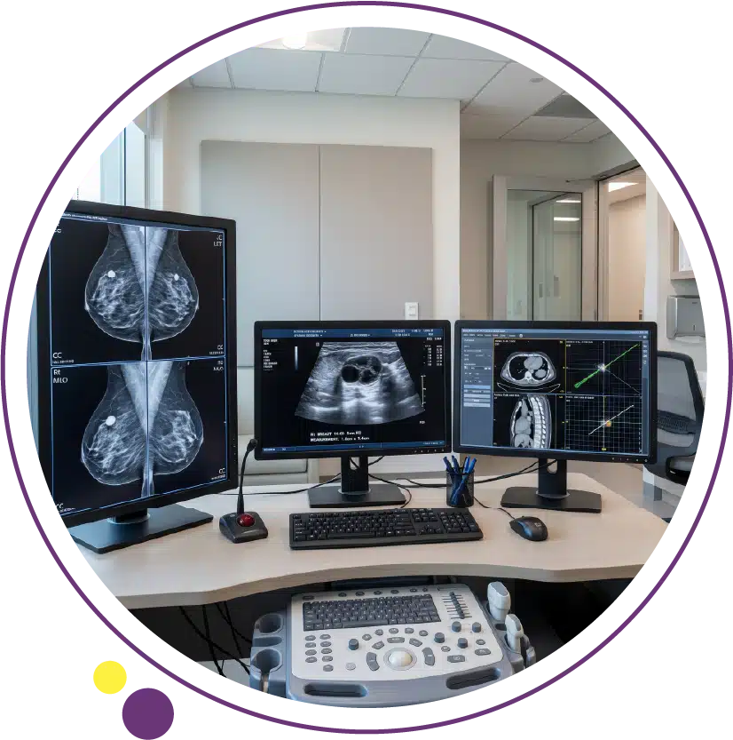
Diagnostic mammogram
A Mammogram used for diagnosis is different from a screening mammogram because it focuses more closely on the area of concern and often includes extra views from different angles. This is commonly the first imaging step after a new symptom or an abnormal screening result. If you want a broader patient-friendly overview before a visit, the older Images article What is a Mammogram fits naturally with this stage of the process.
Breast ultrasound
Ultrasound & Doppler is often used alongside mammography rather than instead of it. A breast ultrasound can help evaluate a lump, clarify an abnormal area, and guide a needle during biopsy when the target is visible on ultrasound. It is also useful because it does not use radiation.
Breast MRI in selected cases
MRI is not needed for every patient with a breast concern, but it can be helpful in selected situations, including certain high-risk patients or cases where mammography and ultrasound do not fully explain a finding. In some practices, Open MRI may also matter for comfort discussions, although breast MRI itself follows a specific protocol and is only used when clinically appropriate.
What does a biopsy involve?
A biopsy removes tissue or cells so a pathologist can determine whether cancer is present. This is the only way to know for sure if a suspicious breast finding is cancer. In modern practice, a needle biopsy is often used first because it is less invasive than surgery and still provides enough tissue to make a diagnosis in many cases. Services and Contact are the most relevant internal pages when patients want to understand what happens after suspicious imaging.
A core needle biopsy is often the preferred approach when breast cancer is suspected because it removes more tissue than fine-needle aspiration without requiring open surgery. Depending on where the abnormality appears best, the biopsy may be guided by ultrasound, mammography, or MRI. At Images, this part of the patient journey usually follows the diagnostic imaging stage rather than replacing it.
If the lesion is visible on ultrasound, Ultrasound & Doppler may pair naturally with an ultrasound-guided biopsy. If it is best seen on mammography, a stereotactic biopsy may be used. If it appears only on MRI, an MRI-guided biopsy may be considered. The guidance method is chosen for accuracy, not because one method automatically means a more serious result.
In many biopsies, a tiny marker clip is placed in the sampled area so it can be identified on future imaging or during treatment planning if needed. This clip is designed to stay in place safely and usually does not cause pain or everyday problems. Some patients also have mild soreness afterward and are advised to avoid strenuous activity for about 24 hours.
What happens after the biopsy?
After tissue is collected, the sample goes to pathology. The pathologist looks at the cells under a microscope and determines whether the tissue is benign, atypical, in situ disease such as DCIS, or invasive cancer. This is the stage where the diagnosis becomes specific, and it is why biopsy results carry more weight than imaging impressions alone. While waiting, many patients find it useful to review the Blog and the Services overview instead of trying to interpret isolated terms online.
If cancer is confirmed, the pathology report often includes more than just the word “cancer.” It may also include tumor type, grade, and biomarker testing. Breast cancers and DCIS are commonly tested for hormone receptors such as ER/PR, because these findings help shape treatment planning.
For invasive cancers, HER2 testing is also important because HER2-positive cancers behave differently and may respond to targeted treatment. Patients do not need to memorize these details before imaging, but it helps to know that diagnosis is not just about finding a mass. It is also about learning what type of cancer it is and what that means for the next step.
How are imaging reports interpreted?
Breast imaging reports often use BI-RADS, which is a standardized system radiologists use to describe findings and recommend what happens next. A BI-RADS category does not replace biopsy, but it helps organize the level of concern and whether the patient needs routine follow-up, short-interval follow-up, or tissue sampling. This is one reason a Mammogram report should be read in context rather than as a standalone answer.
For patients, BI-RADS can reduce confusion because it gives a shared language between radiologists and referring doctors. A suspicious category means the abnormality needs a closer look, not that cancer has already been proven. That distinction matters, and it is one reason diagnostic visits through Images Diagnostic Center are structured around both imaging interpretation and the next practical step.
Are more tests needed after diagnosis?
If a biopsy confirms cancer, some patients need additional imaging or testing before treatment planning is finalized. The exact workup depends on the biopsy result, the size and location of the tumor, lymph node concerns, and whether there are symptoms suggesting disease outside the breast. This is where MRI, CT Scan, or other targeted studies may sometimes enter the discussion, but not every patient needs every scan.
Staging is based on more than imaging alone. Current staging considers tumor size and spread, but also factors such as lymph node involvement, tumor grade, and biomarkers. In practical terms, that means the diagnosis becomes clearer over several connected steps rather than at one single appointment.
How should patients prepare for diagnostic breast imaging?
For a Mammogram, patients are commonly advised not to wear deodorant, talcum powder, or lotion under the arms or on the breasts on the day of the exam, because these can appear on the images. It also helps to mention any new symptoms clearly and bring prior mammograms if they were done elsewhere.
For Ultrasound & Doppler or biopsy appointments, the instructions may vary depending on the planned procedure. In general, patients should tell the team about medications, allergies, pregnancy possibility, and any earlier breast procedures. If biopsy is expected, it is also useful to ask in advance about aftercare and activity limits for the first day.
Planning the right next step in Kuwait
The most useful way to think about breast cancer diagnosis is as a structured pathway, not a single moment. A symptom or abnormal screening result leads to focused imaging, suspicious findings may lead to biopsy, and pathology then clarifies exactly what has been found. That is why patients benefit from choosing the right breast imaging route early through Images Diagnostic Center, Mammogram, Ultrasound & Doppler, or the Contact page.
If you are dealing with a new symptom or an abnormal breast imaging result, checking the nearest branch or contacting the team is the most practical way to understand which diagnostic step should come next.
FAQ
- Is a mammogram enough to diagnose breast cancer?
Not always. A Mammogram can detect suspicious changes, but diagnosis may also need Ultrasound & Doppler and, if concern remains, a biopsy to confirm what the finding actually is.
- Can ultrasound diagnose breast cancer by itself?
Ultrasound & Doppler is very useful for evaluating a breast abnormality, but it does not replace pathology. It can support diagnosis, guide the biopsy, and clarify what the radiologist sees, yet tissue testing is still needed for confirmation.
- Is biopsy always the next step after an abnormal mammogram?
Not always. Sometimes more imaging is needed first, including extra Mammogram views or targeted Ultrasound & Doppler, and some findings are judged safe for short-term follow-up instead of immediate biopsy.
- How long does the diagnosis process usually take?
The timing varies depending on how the abnormality was found, whether extra imaging is needed, and how quickly biopsy and pathology can be arranged. The process often unfolds in stages, so patients should think of diagnosis as a sequence through Services and Contact rather than one single test.
- If the biopsy shows cancer, is that the final report?
It confirms the diagnosis, but more information usually follows. The pathology report may add details about tumor type, grade, and biomarkers such as ER/PR and HER2, which help shape treatment planning.