
An ultrasound scan is one of the most frequently requested imaging tests in clinical practice. It uses high frequency sound waves to create real time images of organs, blood vessels, soft tissues, and developing pregnancies. In Kuwait, physicians commonly request an ultrasound scan as a first line diagnostic tool because it is non ionizing, accessible, and highly effective for many clinical questions.
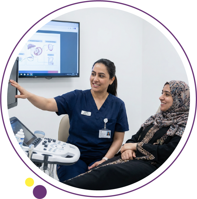
AtImages Diagnostic Center in Kuwait, ultrasound is performed using standardized protocols, structured reporting, and strict patient screening procedures to ensure diagnostic reliability. This article explains how ultrasound works, when it is preferred over other modalities, what its limitations are, the cost of the test, and how patients can prepare properly for the exam.
What is ultrasound scan and how does it work?
An ultrasound scan question usually refers to its basic principle. Ultrasound imaging relies on high frequency sound waves that travel through the body and reflect differently from tissues depending on their density and composition.
A handheld probe called a transducer emits sound waves and receives returning echoes. These echoes are processed by a computer to generate grayscale images in real time. Unlike CT scan in Kuwait or MRI scan in Kuwait, ultrasound does not use ionizing radiation or magnetic fields.
The core technical components include:
- Transducer crystals that convert electrical energy into sound waves
- Echo detection based on tissue impedance differences
- Real time image reconstruction
- Doppler functionality to assess blood flow
Because ultrasound is dynamic, it allows radiologists to assess movement, compressibility, and vascular flow during the examination.
When do doctors typically order an ultrasound scan?
An ultrasound scan is commonly ordered when soft tissue evaluation, fluid assessment, or vascular flow analysis is required.
Typical indications include:
Abdominal ultrasound
- Right upper quadrant pain and suspected gallstones
- Liver Elastography (Ultrasound ) or liver disease assessment
- Kidney stones or hydronephrosis
- Pancreatic inflammation
Pelvic ultrasound
- Ovarian cysts
- Uterine fibroids
- Early pregnancy evaluation
- Pelvic pain
Obstetric ultrasound
- Fetal viability
- Gestational age
- Placental position
- Growth monitoring
Thyroid ultrasound
- Thyroid nodules
- Goiter
- Follow up of suspicious lesions
Musculoskeletal ultrasound
- Tendon injuries
- Joint effusion
- Soft tissue masses
Doppler ultrasound
- Deep vein thrombosis
- Carotid artery stenosis
- Peripheral vascular disease
In many of these scenarios, ultrasound is preferred over CT scan in Kuwait because it avoids radiation exposure and can be repeated safely.
How does Doppler ultrasound assess blood flow?
Doppler ultrasound is a specialized form of vascular ultrasound. It evaluates the frequency shift of sound waves as they bounce off moving red blood cells. This allows visualization and quantification of blood flow direction and velocity.
Clinically, Doppler ultrasound is medically necessary in cases such as:
- Suspected deep vein thrombosis
- Carotid artery plaque evaluation
- Renal artery stenosis
- Varicose vein assessment
- Fetal blood flow monitoring
Color Doppler adds visual mapping of flow direction, while spectral Doppler provides measurable velocity curves. These features help determine whether narrowing, blockage, or abnormal flow patterns are present.
Why is ultrasound often preferred over CT or MRI?
Modality selection depends on the clinical question. An ultrasound scan is often selected first for several reasons:
Advantages over CT scan
- No ionizing radiation
- Bedside availability
- Real time dynamic imaging
- Lower cost in many clinical contexts
Advantages over MRI scan
- Faster setup
- No magnetic safety screening needed
- Better for superficial soft tissue structures
- Real time vascular assessment
However, MRI remains superior for deep soft tissue contrast resolution and neurological evaluation. CT remains the preferred modality for acute trauma and complex internal injuries.
At Images Diagnostic Center, modality selection follows ACR appropriateness criteria to ensure patients receive the most clinically appropriate test.
What are the real limitations of ultrasound imaging?
Although an ultrasound scan is versatile, it has defined limitations:
- Limited penetration in obese patients
- Reduced image clarity through bowel gas
- Operator dependent image acquisition
- Lower sensitivity for deep retroperitoneal structures
- Limited visualization of air filled organs
For example, in suspected appendicitis, ultrasound may be used first, but if visualization is inconclusive, a CT scan in Kuwait may be recommended for definitive evaluation.
Understanding these limitations helps avoid diagnostic delays and ensures appropriate follow up imaging when needed.
Step by step: what happens during an ultrasound scan?
Patients often feel uncertain about what to expect. The process at Images Diagnostic Center follows a structured pathway:
1. Arrival and registration
Patient identification and exam verification are completed.
2. Clinical screening
The technologist reviews:
- Symptoms
- Medical history
- Pregnancy status if relevant
- Previous imaging
3. Preparation
Depending on the exam type:
- Fasting may be required for abdominal ultrasound
- Full bladder may be needed for pelvic ultrasound
4. Scanning procedure
- Gel is applied to improve sound transmission
- The probe is moved over the area of interest
- Real time images are captured
- Doppler settings are activated if vascular assessment is required
5. Post scan
There is no recovery period. Patients may resume normal activity immediately unless a biopsy was performed.
How should patients prepare for different types of ultrasound?
Preparation improves diagnostic accuracy.
Abdominal ultrasound preparation
- Fast for 6 to 8 hours
- Avoid fatty meals prior to exam
Pelvic ultrasound preparation
- Drink water to achieve a full bladder
- Do not urinate before scan
Renal Doppler ultrasound
- Light fasting may be advised
- Avoid heavy meals
No preparation is required for thyroid, musculoskeletal, or vascular limb ultrasound in most cases.
How accurate is an ultrasound scan compared to other imaging tests?
Accuracy depends on the clinical indication and operator expertise.
For gallstones, ultrasound sensitivity exceeds 90 percent. For deep abdominal masses, MRI scans in Kuwait may provide superior soft tissue contrast. For bone fractures, X-ray in Kuwait remains first line.
Diagnostic confidence in ultrasound depends on:
- Proper probe selection
- Adequate patient preparation
- Absence of interfering bowel gas
- Experienced sonographers
- Radiologist interpretation
Structured reporting enhances clarity and supports referring physicians in decision making.
Can ultrasound detect cancer?
Ultrasound can detect suspicious masses but cannot confirm malignancy alone. For example:
- Thyroid nodules are categorized based on risk patterns
- Breast imaging may include ultrasound as an adjunct to mammography
- Liver lesions may require Cardiac MRI or CT Scan for characterization
In breast imaging, standardized BI RADS assessment is used when appropriate. If imaging suggests malignancy, biopsy under ultrasound guidance may be performed.
Is ultrasound safe for pregnancy?
Yes. An ultrasound scan does not use ionizing radiation and is considered safe during pregnancy when performed for medical indications.
Obstetric ultrasound helps assess:
- Fetal heartbeat
- Growth parameters
- Placental position
- Amniotic fluid levels
Professional guidelines recommend limiting exposure to medically necessary indications and avoiding non diagnostic scanning.
How long does it take to receive ultrasound scan results?
Radiology report turnaround time depends on exam complexity and clinical urgency.
Factors influencing reporting include:
- Case complexity
- Need for correlation with previous imaging
- Requirement for Doppler analysis
- Radiologist workload
At an accurate diagnosis imaging center in Kuwait, structured workflow and standardized templates support timely and clinically precise reporting.
Ultrasound guided procedures
Ultrasound is frequently used to guide minimally invasive procedures, including:
- Thyroid fine needle aspiration
- Breast biopsy
- Joint aspiration
- Abscess drainage
Real time visualization increases procedural accuracy and reduces complications compared to blind techniques.
After ultrasound guided biopsy:
- Mild soreness may occur
- Patients should monitor for swelling or bleeding
- Follow up instructions are provided
How does ultrasound compare to bone density and breast imaging?
Ultrasound cannot replace DEXA Scan for osteoporosis evaluation. DEXA Scan measures bone mineral density using low dose X-ray technology.
For breast cancer screening, Mammogram remains the standard screening modality. Ultrasound is complementary, particularly in dense breast tissue or when evaluating palpable lumps.
Understanding modality strengths prevents inappropriate substitution.
Clinical quality standards in ultrasound imaging
Accurate ultrasound imaging depends on adherence to recognized standards such as:
- ACR appropriateness guidelines
- Radiation avoidance principles
- Structured reporting protocols
- Quality control of equipment
- Regular technologist training
Consistency in technique improves reproducibility and reduces variability.
When should ultrasound not be the first imaging test?
In certain scenarios, other modalities are preferred:
- Major trauma requiring CT scan
- Complex neurological conditions requiring MRI scan
- Evaluation of pulmonary embolism with Cardiac CT
- Detailed musculoskeletal ligament evaluation with 3 Tesla MRI
Appropriate imaging selection improves diagnostic clarity and reduces unnecessary repeat testing.
Conclusion
An ultrasound scan remains one of the most versatile and safe diagnostic tools in modern radiology. Its ability to provide real time visualization without radiation makes it a preferred first line investigation for many abdominal, pelvic, vascular, and soft tissue conditions.
At Images Diagnostic Center in Kuwait, ultrasound imaging is integrated into a broader diagnostic framework that includes MRI scan, CT scan, mammogra, X-ray, and DEXA Scan. Each modality is selected based on clinical indication, safety considerations, and diagnostic appropriateness.
Understanding how ultrasound works, when it is preferred, and where its limitations lie allows patients and referring clinicians to make informed decisions. Imaging delivers the greatest value when selected thoughtfully, performed accurately, and interpreted within proper clinical context.