
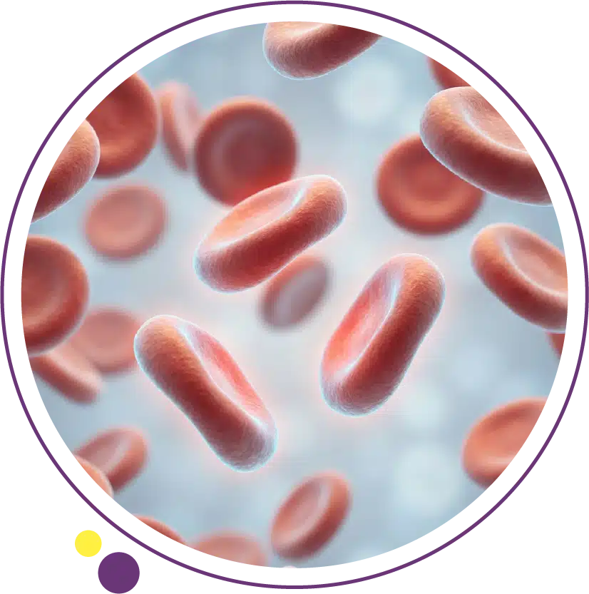
Blood cancers, such as leukemia, lymphoma, and multiple myeloma, often begin quietly. Unlike solid tumors that may form a visible mass, these diseases develop within the bone marrow, lymphatic system, and bloodstream, meaning early changes can be subtle, confusing, and easily mistaken for everyday fatigue or infection.
Yet the earlier blood cancer is detected, the greater the opportunity for precise diagnosis, personalized treatment planning, and improved long-term outcomes.
At Images Diagnostic Center in Kuwait, advanced diagnostic imaging plays a critical role in supporting physicians when blood cancer is suspected.
Technologies such as 3 Tesla MRI scans, high-resolution CT imaging, ultrasound and Doppler studies, and bone density (DEXA) scanning provide clinicians with the anatomical and functional detail needed to assess disease spread, organ involvement, and treatment response.
This article explains seven early warning signs of blood cancer and how medical imaging in Kuwait supports accurate clinical evaluation, staging, and monitoring when these symptoms appear.
Understanding Blood Cancer and Why Imaging Matters
Blood cancers arise from abnormal production and function of blood cells. Instead of healthy white cells that protect against infection, cancerous cells multiply uncontrollably, crowding out normal blood cells and disrupting immune and clotting functions. Depending on the type, blood cancer may affect the bone marrow, lymph nodes, spleen, liver, or other organs.
Laboratory blood tests are usually the first step in diagnosis, but imaging adds critical information. Advanced MRI for accurate diagnosis, CT scans for lymph node and organ assessment, and ultrasound for soft tissue evaluation allow doctors to visualize where disease is present, how far it has progressed, and how the body is responding to therapy.
In Kuwait, Images Diagnostic Center provides physicians and patients with comprehensive, accredited radiology services designed to deliver reliable, reproducible, and clinically meaningful results.
1. Persistent Fatigue That Does Not Improve with Rest
One of the earliest and most overlooked symptoms of blood cancer is persistent fatigue. This is not ordinary tiredness; it is a deep exhaustion that does not resolve after sleep or rest.
Blood cancer interferes with red blood cell production, leading to anemia. When tissues receive less oxygen, patients experience weakness, shortness of breath, and reduced exercise tolerance.
While blood tests detect anemia, imaging helps explain its cause. A 3 Tesla MRI scan in Kuwait can evaluate bone marrow activity, revealing abnormal cellular infiltration that may be suppressing healthy blood cell production. In some cases, MRI also shows bone marrow edema or replacement that points toward leukemia or myeloma.
2. Unexplained Weight Loss and Reduced Appetite
Unexpected weight loss, especially when combined with reduced appetite or early fullness, may indicate involvement of internal organs such as the spleen or liver. In blood cancers like lymphoma or leukemia, these organs may enlarge as they filter abnormal cells from the bloodstream.
A CT scan in Kuwait provides a detailed map of abdominal organs and lymph nodes, allowing doctors to identify enlargement, infiltration, or compression of surrounding tissues. Advanced CT imaging protocols at Images Diagnostic Center enable accurate measurement of organ size and detection of subtle changes that may not be felt on physical examination.
What makes 3 Tesla MRI more accurate in blood cancer evaluation?
A 3 Tesla MRI produces a magnetic field twice as strong as conventional MRI systems. This higher field strength translates into improved signal-to-noise ratio and sharper images, which is particularly valuable when examining bone marrow, spine, brain, and soft tissues affected by blood cancers.
In multiple myeloma, for example, advanced MRI for accurate diagnosis can detect tiny marrow lesions before they become visible on X-ray or CT. This early detection helps oncologists classify disease stage, predict fracture risk, and select the most appropriate therapy.
3. Frequent Infections or Slow Recovery
Because blood cancers disrupt white blood cell function, patients often develop repeated infections, slow wound healing, or persistent fevers. These signs reflect immune system compromise.
When infection is suspected in organs such as the lungs, sinuses, or abdomen, CT imaging provides fast, high-resolution views that guide treatment decisions. At Images Diagnostic Center, CT scans in Kuwait are performed using optimized dose protocols to ensure diagnostic clarity while minimizing radiation exposure.
4. Swollen Lymph Nodes in the Neck, Armpits, or Groin
Lymph nodes act as filters for immune cells, and they often enlarge when abnormal lymphocytes accumulate. Painless swelling in the neck, underarms, or groin is a common early sign of lymphoma.
Ultrasound and Doppler imaging are frequently used to evaluate superficial lymph nodes. These techniques show node size, internal structure, and blood flow patterns, helping doctors differentiate between reactive (infection-related) and malignant enlargement. When deeper nodes are involved, CT scans or MRI provide comprehensive mapping for staging and biopsy planning.
5. Bone Pain or Increased Fracture Risk
Bone pain, particularly in the back, ribs, or hips, may signal bone marrow infiltration or bone weakening caused by multiple myeloma. Cancerous plasma cells can erode bone, leading to fractures even with minor stress.
Bone density testing (DEXA scan) measures mineral content and identifies early bone loss. MRI scans reveal marrow lesions, spinal compression fractures, and nerve involvement. Together, these tools guide treatment decisions, including chemotherapy, radiation, or orthopedic support.
6. Night Sweats and Persistent Fever
Unexplained fever and drenching night sweats are classic “B-symptoms” in lymphoma and advanced leukemia. They reflect the body’s inflammatory response to cancer cells.
CT imaging of the chest, abdomen, and pelvis is often used to look for enlarged lymph nodes, organ involvement, or hidden infections. Advanced CT imaging in Kuwait at Images Diagnostic Center allows rapid whole-body assessment when these systemic symptoms appear.
How fast can CT scan results be obtained for suspected blood cancer?
In urgent clinical situations, such as suspected lymphoma with organ compression or infection, CT scan results are typically available within hours. At Images Diagnostic Center, most CT imaging reports are interpreted promptly by board-certified radiologists, allowing physicians to make timely diagnostic and treatment decisions.
7. Unusual Bruising or Bleeding
Low platelet levels in blood cancer can cause easy bruising, nosebleeds, or bleeding gums. While laboratory tests identify platelet abnormalities, imaging may reveal internal bleeding or organ involvement that explains these changes.
Ultrasound and CT scans can detect splenic enlargement or abdominal bleeding, supporting a complete clinical assessment.
The Role of Imaging in Blood Cancer Diagnosis and Monitoring
Medical imaging is not only about finding disease; it is also about understanding how it behaves over time. Advanced MRI, CT scans, and ultrasound are used throughout a patient’s journey to:
- Establish the extent of disease at diagnosis
- Guide biopsies and treatment planning
- Monitor response to chemotherapy or radiation
- Detect early relapse or complications
At Images Diagnostic Center in Kuwait, every scan is performed using evidence-based protocols and interpreted by experienced radiologists who specialize in oncologic imaging.
Comparing Key Imaging Modalities in Blood Cancer Care
| Imaging Modality | Primary Clinical Use | Why It Matters in Blood Cancer |
| 3 Tesla MRI scan in Kuwait | Bone marrow, spine, brain, soft tissue | Detects early marrow infiltration and nerve involvement |
| CT scan | Chest, abdomen, pelvis | Identifies lymph node enlargement and organ involvement |
| Ultrasound & Doppler | Superficial lymph nodes, organs | Differentiates benign vs malignant node changes |
| DEXA Scan | Bone density | Assesses fracture risk in myeloma and chronic leukemia |
| Mammography | Breast tissue | Evaluates secondary or coincidental breast findings |
Home Radiology for Patients with Limited Mobility
Some patients with advanced blood cancer or post-treatment weakness cannot easily travel to a medical center. Home radiology service in Kuwait, known as Images GO, brings mobile imaging for patients at home using certified portable equipment. X-rays and ultrasound exams are performed by trained technologists, and images are reviewed by the same expert radiologists who interpret in-center scans.
This service supports continuity of care while reducing infection risk and physical strain.
Why Accreditation Matters in Cancer Imaging
When imaging influences life-changing decisions, quality standards are essential. Images Diagnostic Center holds both American College of Radiology (ACR) accreditation and Accreditation Canada Platinum status. These certifications confirm that equipment, protocols, safety measures, and professional expertise meet rigorous international benchmarks.
For patients facing possible blood cancer, this means every MRI scan, CT scan, and ultrasound study is performed with precision, consistency, and accountability.
Conclusion
Recognizing blood cancer symptoms early can make a meaningful difference in diagnosis and treatment. Persistent fatigue, unexplained weight loss, swollen lymph nodes, bone pain, and unusual bleeding are signals that deserve careful medical evaluation.
Advanced imaging in Kuwait, particularly 3 Tesla MRI scans, high-resolution CT imaging, and expert ultrasound diagnostics, provides the clarity physicians need to confirm findings, stage disease, and monitor progress.
At Images Diagnostic Center, patients benefit from internationally accredited technology, subspecialty radiologists, and compassionate care designed to support accurate, timely diagnosis. Proactive imaging, combined with laboratory testing and clinical expertise, remains one of the most powerful tools for protecting health and guiding effective cancer care.
FAQs
- What blood tests are usually ordered before imaging for suspected blood cancer?
Doctors typically request a complete blood count, peripheral smear, and sometimes specific tumor markers before recommending imaging.
- Can MRI replace a bone marrow biopsy?
MRI provides valuable information about marrow involvement, but biopsy is still required for definitive diagnosis.
- Is CT scanning safe for patients undergoing chemotherapy?
Yes, when medically indicated and performed with dose-optimized protocols, CT scans are safe and clinically important.
- How often is imaging repeated during blood cancer treatment?
Frequency depends on cancer type, treatment plan, and clinical response, ranging from every few weeks to several months.
- Can ultrasound detect leukemia?
Ultrasound does not diagnose leukemia directly but helps assess organs and lymph nodes affected by the disease.
- Does home radiology provide the same quality as in-center scans?
Mobile imaging is ideal for follow-up X-rays and ultrasound, while MRI and CT require specialized in-center equipment.
Book your diagnostic imaging with confidence
- 3 Tesla MRI scans
- CT scans and cardiac CT imaging
- Ultrasound & Doppler studies
- Bone density (DEXA) scans
- Home radiology services (Images GO)
Contact Images Diagnostic Center
📞 (+965) 1899 888
Accurate diagnosis begins with precise imaging. Schedule your consultation or imaging study at Images Diagnostic Center and take a proactive step toward informed healthcare decisions.