
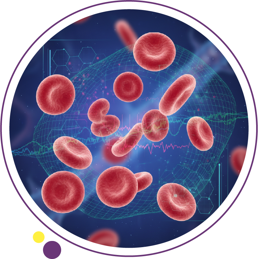
Blood cancer types are usually grouped into three main families: leukemia, lymphoma, and myeloma. These cancers all affect blood, bone marrow, or the lymphatic system, but they do not begin in the same cells or follow the same diagnostic pathway. At Images Diagnostic Center, that difference matters because the role of CT Scan, MRI, Ultrasound & Doppler, or Digital X-ray depends on the suspected disease and the reason for referral.
Many patients search this topic because they want a simple explanation of the main categories before they face blood tests, biopsy results, or imaging referrals. The broad answer is simple, but the details matter: leukemia is mainly a bone marrow and blood-cell cancer, lymphoma begins in lymphocytes and the lymphatic system, and myeloma starts in plasma cells. In this article, we will discuss the main blood cancer types, how they differ, what symptoms can overlap, and how doctors use testing and imaging to tell them apart.
What are the main blood cancer types?
Blood cancers develop when blood cells or related cells begin to grow abnormally. According to major cancer references, they can begin in the bone marrow, where blood cells are made, or in the lymphatic system, which helps fight infection. That is why a patient referred through our Services pathway may need different tests depending on whether the concern is marrow-based, lymph-node based, or linked to plasma cells.
The three main groups are leukemia, lymphoma, and myeloma, but each group contains many subtypes. Leukemia can be acute or chronic and can arise from different blood cell lines. Lymphoma is commonly divided into Hodgkin and non-Hodgkin lymphoma. Myeloma also has its own diagnostic and treatment pathway, especially because it is closely tied to plasma cells and bone marrow function. That is why the exact subtype matters just as much as the broad category when patients are moving from consultation to Contact and imaging planning.
Leukemia
Leukemia is a broad term for cancers of the blood cells. The type of leukemia depends on which blood cell becomes cancerous and whether it grows quickly or slowly, which is why doctors often describe it as acute or chronic, and as myeloid or lymphoid. At Images Diagnostic Center, this matters because imaging is usually supportive in leukemia, while blood tests and bone marrow analysis remain central to diagnosis.
Acute leukemias tend to grow faster and often cause symptoms that develop over weeks rather than years. Chronic leukemias may be found more slowly, and some are discovered after routine blood work rather than a dramatic symptom. Patients who are trying to understand the next step after abnormal lab results can review our Services page first, then move to the right imaging exam only when it has a clear clinical purpose.
Common leukemia symptoms can include fatigue, fever, night sweats, loss of appetite, bruising, bleeding, or infections that do not settle normally. These symptoms are not unique to leukemia, but when they appear together or continue, doctors investigate further with blood work and sometimes additional studies. If symptoms also raise concern about organs, chest findings, or complications, our CT Scan or MRI services may become part of the wider workup.
Lymphoma
Lymphoma begins in lymphocytes, which are white blood cells that help the body fight infection. This type of blood cancer is closely tied to the lymphatic system, so it often raises questions about enlarged lymph nodes, swelling, or deeper disease in the chest, abdomen, or pelvis. That is why referrals involving suspected lymphoma often lead more directly to CT Scan or sometimes MRI than many leukemia workups do.
The two broad lymphoma groups are Hodgkin lymphoma and non-Hodgkin lymphoma. That sounds simple, but non-Hodgkin lymphoma alone includes many subtypes, some slow-growing and some aggressive. Patients who want to understand why different protocols may be used during staging can also read our previous Images article on types of CT scan alongside the main CT Scan service page.
Lymphoma symptoms often include enlarged lymph nodes, fever, drenching night sweats, and unexplained weight loss. Those are sometimes called B symptoms, and they matter because they can influence how doctors think about disease activity and staging. If a patient has a lump, swelling, or abdominal symptoms, our Ultrasound & Doppler service may also help in selected cases, especially when the question involves superficial nodes or abdominal organs.
Radiology references note that if lymphoma is diagnosed, doctors may use chest X-ray, body CT, body MRI, or abdominal ultrasound to look for enlarged lymph nodes and to assess spread. This is one of the clearest examples of how imaging supports blood cancer care without replacing pathology. At Images Diagnostic Center, the imaging choice depends on the referral question, the body area, and the information the treating team still needs.
Myeloma
Myeloma starts in plasma cells, which are immune cells normally found in the bone marrow. This makes myeloma different from leukemia and lymphoma in both symptoms and imaging needs. Because it is closely linked to marrow activity and bone involvement, patients with suspected or confirmed myeloma are more likely to need MRI, Digital X-ray, or sometimes CT Scan as part of their evaluation.
Common myeloma symptoms include bone pain, marked tiredness, shortness of breath, muscle weakness, increased thirst, and unintentional weight loss. Another important point is that myeloma does not always cause symptoms early, and some people first learn about it after blood tests performed for another reason. That is why our Services and Contact pages are most useful when they are part of a doctor-led plan rather than a self-diagnosis route.
What does “and more” mean?
For SEO, many articles simplify the topic to three main blood cancer types, and that is still the clearest place for most readers to start. But some trusted patient organizations also include related groups such as myelodysplastic syndromes, or MDS, and myeloproliferative neoplasms, or MPNs, under the wider blood cancer umbrella. That is part of the reason the title often says “leukemia, lymphoma & more,” even though the main educational focus usually stays on the big three. Our Blog is useful here because it helps patients move from broad terms to the specific question their doctor is actually investigating.
This wider classification also explains why some disease names sound unfamiliar at first. A patient may hear terms such as Hodgkin lymphoma, diffuse large B-cell lymphoma, AML, CLL, or MDS long before they understand how those terms fit together. At Images Diagnostic Center, we try to keep the diagnostic journey clearer by connecting the right imaging service, such as Open MRI or Ultrasound & Doppler, to the actual referral question rather than to a generic cancer label.
How doctors tell blood cancer types apart
The first step is usually not imaging alone. Doctors begin with symptoms, physical examination, and blood tests, then decide whether a biopsy or bone marrow test is needed. If the concern points toward lymphoma, imaging may be used earlier to map lymph nodes and organ involvement. If the concern is leukemia or myeloma, lab patterns and marrow findings usually drive the diagnosis first, while MRI, CT Scan, or Digital X-ray answer more specific follow-up questions. (Radiologyinfo.org)
A bone marrow biopsy is especially important when doctors need to confirm marrow-based disease, identify the abnormal cells, and define the subtype more clearly. That is one reason imaging should never be presented as a stand-alone way to diagnose all blood cancers. At Images Diagnostic Center, our role is to support that pathway when imaging is the right next step, not to replace hematology evaluation. (Radiologyinfo.org)
Imaging becomes particularly useful when the question involves lymph nodes, organ enlargement, chest symptoms, abdominal disease, or bone pain. That is why the same patient may need very different imaging depending on whether the suspected type is lymphoma, myeloma, or a complication related to leukemia. Our Services page helps patients understand these differences before they choose a branch or discuss the right exam with the team. (Radiologyinfo.org)
Symptoms that can overlap across blood cancer types
One reason this topic is confusing is that the symptoms often overlap. Fatigue, weight loss, fever, night sweats, bruising, bleeding, infections, swollen nodes, and bone pain can all appear in blood cancer, but not every symptom points to the same disease. That is why persistent symptoms should be assessed by a doctor first, and why our Contact page is most useful after there is a clear referral question.
Lymphoma often stands out because of swollen lymph nodes and classic B symptoms. Leukemia often raises concern when infections, bruising, bleeding, or fatigue are linked to abnormal blood counts. Myeloma more often brings bone pain or unexplained weakness into the picture. These are patterns, not guarantees, which is why patients in Kuwait often move through several steps before reaching the right Services pathway at Images Diagnostic Center.
Choosing the next step in Kuwait
The most practical way to understand blood cancer types is to focus on where the abnormal cells start and what that means for diagnosis. Leukemia, lymphoma, and myeloma can sound similar at first, but they create different patterns of symptoms and often lead to different testing pathways. If your doctor has already suggested imaging, our Services, Contact, and scan-specific pages can help you prepare for the right next step.
If you want, I can also turn this into a stricter publish-ready version with even denser internal linking and no citations showing in the body.
FAQ
- Which are the three main blood cancer types?
The three main groups are leukemia, lymphoma, and myeloma. Each starts in a different kind of cell, which is why patients may need different blood tests, biopsy methods, or imaging through our Services pages.
- Is lymphoma the same as leukemia?
No. Leukemia mainly involves blood-forming cells in the blood and bone marrow, while lymphoma begins in lymphocytes and commonly affects lymph nodes and the lymphatic system. That difference is one reason CT Scan is often more central in lymphoma staging than in many straightforward leukemia evaluations.
- Is myeloma a type of blood cancer?
Yes. Myeloma is a blood cancer because it starts in plasma cells, which are immune cells found in bone marrow. Because bone involvement is common, our MRI and Digital X-ray services may be relevant when a doctor is evaluating pain or structural complications.
- Can imaging alone tell which blood cancer type someone has?
Usually no. Imaging can show enlarged lymph nodes, organ changes, or bone-related findings, but blood tests, pathology, and sometimes marrow studies are still needed to define the exact disease. That is why our CT Scan, MRI, and Ultrasound & Doppler services support diagnosis rather than replace it.
- What does “and more” include in blood cancer articles?
It usually means there are other blood-cancer-related groups beyond the main three, including MDS and MPNs, plus many subtypes inside leukemia, lymphoma, and myeloma themselves. If your referral includes an unfamiliar term, our Blog and Contact pages can help you navigate the next step more clearly.